Locations:

First study to assess sublingual blood flow during SML
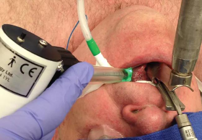
Image content: This image is available to view online.
View image online (https://assets.clevelandclinic.org/transform/fd107da6-6018-4bc7-b62a-59bc8643d502/15-ENT-2834-Bryson-Consult-QD-Image-1_jpg)
15-ENT-2834-Bryson-Consult-QD-Image
Advertisement
Cleveland Clinic is a non-profit academic medical center. Advertising on our site helps support our mission. We do not endorse non-Cleveland Clinic products or services. Policy
Surface capillaroscopy is a novel method of evaluating microcirculation during microlaryngoscopic procedures. Impaired microcirculation during surgery can lead to local tissue compromise and postoperative complications. Yet despite the widespread use of suspension microlaryngoscopy (SML), no real-time measures of tongue perfusion have been quantified. In fact, until the Head & Neck Institute completed its recent study, surface capillaroscopy had never before been used to assess tongue and sublingual circulation during SML.
Our prospective study of surface capillaroscopy involved the use of laser Doppler technology to evaluate tongue perfusion during SML. Our study population was made up of 15 adults who were undergoing SML for various reasons.
Perfusion was assessed in a series of 20-second video recordings to examine the sublingual microcirculation. Recordings were made prior to scope insertion, immediately following suspension, at regular intervals throughout the procedure (for patients who were undergoing longer surgeries) and finally when the scope was removed (Figure 1). The duration of surgery ranged from 15 to 80 minutes. Following surgery, patients completed a questionnaire designed to determine the extent of postoperative complications, if any.
Intraoperative analysis of the videos revealed that microvascular flow indices had decreased significantly during all procedures. The longer surgeries were associated with longer periods of decreased flow, although some improvement in flow was observed as the procedure progressed.
Advertisement
The reductions in perfusion appeared to correlate with postoperative oropharyngeal complications. Diminished perfusion appeared to be associated with larger laryngoscopes, elevated vector suspensions, difficult microsurgical exposure and longer procedures.
Given these preliminary findings, it may be worthwhile to consider periodic removal from suspension in an effort to limit side effects, and surface capillaroscopy allows us to determine when to do so.
Decreased perfusion during SML can lead to postoperative oropharyngeal complications such as taste disturbance and tongue numbness or weakness secondary to lingual, hypoglossal and glossopharyngeal nerve injuries. While these complications are often minor and temporary, they can cause distress while they persist. Knowing the extent of the loss of perfusion allows us to determine whether we should ease off suspension intraoperatively until blood flow is restored.
The capillaroscopy cameras that we use obtain orthogonal polarization spectral images with green light at 550 nm. This wavelength this preferentially absorbed by oxyhemoglobin in red blood cells and, with filtration, it allows us to clearly visualize the microcirculation.
We conclude that surface capillaroscopy is a safe and easily employed technology to evaluate sublingual and tongue perfusion during SML. This technology will allow for further study of the impact of microcirculatory changes during SML on a number of variables and outcomes. Future studies can be aimed at determining the ideal timing for this strategy.
Advertisement
Dr. Bryson is Director of the Voice Center and Section Head of Laryngology in the Head & Neck Institute. He can be reached at 216.445.6468 or brysonp@ccf.org
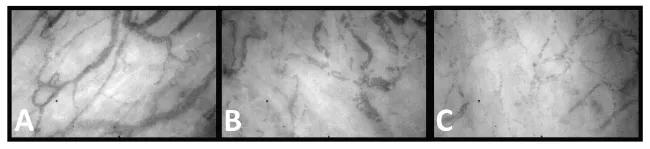
Image content: This image is available to view online.
View image online (https://assets.clevelandclinic.org/transform/73214987-c578-4038-8eb6-33e83b19deb1/15-ENT-2834-Byson-Consult-QD-Inset-Image_jpg)
Figure 1. These CapiScope® images show the microcirculation in the sublingual/ventral tongue mucosa during microlaryngoscopy. A: Prior to laryngoscope insertion and fixation, flow is normal in both capillaries (1 RBC thick) and venules (> 1 RBC thick). B: Ten minutes postsuspension, the capillaries exhibit sluggish to absent flow. Stasis is noted, and venules are congested. C: Image shows the return of capillary blood flow, although it remains sluggish compared with the presuspension flow. (The image frame is approximately 700 × 450 μm.)
Advertisement
Advertisement

With a wide scope of skills, comprehensive otolaryngologists care for patients of all ages in the community
Research on children with UHL explores the quality-of-life benefits and outcomes of cochlear implants
A look at how custom-fitted oral appliances work and when they’re a good fit for patients
Subtle information gleaned from clinical examinations prompted concern
A new single-port system well-suited for oropharyngeal cancer treatment

Challenging case requires outside-the-box approach
Collaborative and multidisciplinary approach necessary for treatment

The tri-vector gracilis procedure uses a thin muscle from the thigh to help create a natural mimetic smile