Locations:

Report details imaging benefit, limitations
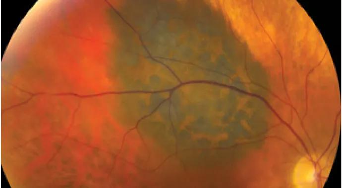
Image content: This image is available to view online.
View image online (https://assets.clevelandclinic.org/transform/9da0a3f5-9ef4-4dcd-9cd8-a59fed562766/Singh-image-690-x-380_jpg)
Singh-image-690-x-380
Optical coherence tomography (OCT) has become essential to the practice of ophthalmology, offering superior resolution of the retina and some of the adjacent tissues for diagnosis and treatment of eye diseases.
Advertisement
Cleveland Clinic is a non-profit academic medical center. Advertising on our site helps support our mission. We do not endorse non-Cleveland Clinic products or services. Policy
New OCT modalities have now been assimilated into ophthalmic oncology to help track the status of ocular and periocular tumors through retinal symptoms.
“Optical coherence tomography imaging of ocular and periocular tumours,” a review of newer OCT modalities including anterior segment (AS), ultra-high resolution (UHR), spectral domain (SD) and enhanced depth imaging (EDI), describes the current advantages and limitations of their use.
The paper also offers examples of how these advanced OCT modalities have been used, with mixed results, in the diagnosis and treatment of skin cancers of the eyelid, ocular surface lesions, iris and ciliary body tumors, nevus and melanoma, lymphoma, hemangioma, metastasis and retinoblastoma.
In one study involving 34 eyes of 34 patients with conjunctival lesions believed to be ocular surface squamous neoplasia (OSSN) and pterygia, for example, UHR-OCT revealed significant morphologic correlation with biopsy results.
EDI-OCT has been a helpful tool in identifying potentially important signs that can help differentiate common choroidal naevus from the much less common but potentially fatal melanoma. “Subretinal fluid (91 percent versus 14 percent), retinal odema (61 percent versus 14 percent) and subretinal deposits (61 percent versus 11 percent) all have significantly higher prevalence in melanoma compared to naevus,” the paper reports.
Even in eye-related diseases such as lymphoma, for which biopsy remains the “gold standard,” OCT has been shown to be helpful in the diagnosis. In a study of 61 eyes with biopsy-proven vitreoretinal lymphoma, OCT images showed nodular hyper-reflective lesions at the level of the retinal pigment epithelium (RPE). Other studies have shown the presence of hyper-reflective bands or nodules distributed across the retinal layers.
Advertisement
“(OCT) is not going to become the gold standard (for ophthalmic oncology),” says Arun Singh, MD, Director of the Department of Ophthalmic Oncology in the Cole Eye Institute at Cleveland Clinic and the lead author of the review. “As of now, I don’t see that. Diagnosis is (still) the sum total of examination and tests, with a reliance more on exams than tests.”
Yet, he adds, OCT “can give us a lot of useful information (on tumors) in reference to what the tumor is doing to the retina. Tumors are typically under the retina. And under the retina they don’t show very well. But, OCT can tell us whether there is the presence or absence of subretinal fluid, the presence or absence of radiation damage to the retina.”
“Even with limitations, the paper highlights many things we can figure out, such as whether the tumor is benign or malignant, or growing or not growing based on what it does (in the area) around itself, meaning the retina and the pigment epithelium,” Dr. Singh adds.
The new imaging modalities offer improvements in diagnosis, treatment planning and monitoring response in patients with ophthalmic tumors,” the paper says.
“It is important, however, to understand the limitations of OCT, mainly its penetration in deep pigmented tissue. Whereas imaging of the cornea is mainly limited by resolution, imaging of the skin and uvea is limited by penetration… Imaging of pigmented and vascularized lesions is also limited.
“OCT provides valuable information regarding the status of the retina, RPE and inner choiroid, but ultrasonography including UBM is still essential in the majority of patients with intraocular tumours.”
Advertisement
Advertisement

First-of-its-kind research investigates the viability of standard screening to reduce the burden of late-stage cancer diagnoses

Global R&D efforts expanding first-line and relapse therapy options for patients

Study demonstrates ability to reduce patients’ reliance on phlebotomies to stabilize hematocrit levels

A case study on the value of access to novel therapies through clinical trials

Findings highlight an association between obesity and an increased incidence of moderate-severe disease

Cleveland Clinic Cancer Institute takes multi-faceted approach to increasing clinical trial access 23456
Key learnings from DESTINY trials

Overall survival in patients treated since 2008 is nearly 20% higher than in earlier patients