Locations:

Complete resection of tumor cells requires novel reconstruction
By Nathan Mesko, MD and Joshua Lawrenz, MD
Advertisement
Cleveland Clinic is a non-profit academic medical center. Advertising on our site helps support our mission. We do not endorse non-Cleveland Clinic products or services. Policy
Complete resection is the gold standard for minimizing the chance of local recurrence in primary bone sarcoma. Sometimes, in an effort to resect without leaving behind any tumor cells, large voids can be created, making functionality impossible without reconstruction. But bone and soft tissue tumors often present ways that make routine resection and reconstruction impossible. Creative thinking can improve the chances of limb salvage over an amputation alternative, and can determine long- and short-term reconstruction success.
These slides demonstrate the case of a 70-year-old gentleman who presented with four years of progressively worsening left thigh cramping, with no specific history of trauma. Just prior to presentation, his pain progressed to the point where he stated the leg “felt like it was going to break.”

Image content: This image is available to view online.
View image online (https://assets.clevelandclinic.org/transform/860e6e39-76d0-411b-bc3b-7ed59ea43d95/16-ORT-2502-Mesko-Slide-01-150x103_jpg)
Advertisement

Image content: This image is available to view online.
View image online (https://assets.clevelandclinic.org/transform/09544417-2c9f-4012-86cf-aaa4c21da080/16-ORT-2502-Mesko-Slide-02-150x103_jpg)

Image content: This image is available to view online.
View image online (https://assets.clevelandclinic.org/transform/887cf655-f282-4f2b-8d9a-1a89a3253ac1/16-ORT-2502-Mesko-Slide-03-150x103_jpg)

Image content: This image is available to view online.
View image online (https://assets.clevelandclinic.org/transform/4034a48f-6323-4c3c-972e-c9dbdf0822f8/16-ORT-2502-Mesko-Slide-04-150x103_jpg)

Image content: This image is available to view online.
View image online (https://assets.clevelandclinic.org/transform/34df3449-5e36-47ec-88dc-cf0f78ef5620/16-ORT-2502-Mesko-Slide-05-150x103_jpg)
Advertisement

Image content: This image is available to view online.
View image online (https://assets.clevelandclinic.org/transform/03c5d966-23dc-4f92-b10b-f49827d8a5aa/16-ORT-2502-Mesko-Slide-06-150x103_jpg)

Image content: This image is available to view online.
View image online (https://assets.clevelandclinic.org/transform/9ff365a5-019e-4a24-9270-989a5e2df464/16-ORT-2502-Mesko-Slide-07-150x103_jpg)

Image content: This image is available to view online.
View image online (https://assets.clevelandclinic.org/transform/1a74e5f3-906c-4950-922f-54da8372741a/16-ORT-2502-Mesko-Slide-08-150x103_jpg)

Image content: This image is available to view online.
View image online (https://assets.clevelandclinic.org/transform/6e1e5a28-0dbb-45df-833d-7afbc50d7126/16-ORT-2502-Mesko-Slide-09-150x103_jpg)

Image content: This image is available to view online.
View image online (https://assets.clevelandclinic.org/transform/62f22d85-f795-4837-bd05-44ce79a9ede1/16-ORT-2502-Mesko-Slide-10-150x103_jpg)
Advertisement

Image content: This image is available to view online.
View image online (https://assets.clevelandclinic.org/transform/92622ff9-047d-4292-97cc-bd3a4c683bdd/16-ORT-2502-Mesko-Slide-11-150x103_jpg)
Slide 1/11
Dr. Mesko is Director of the Musculoskeletal Tumor Center. Dr. Lawrenz is a resident in orthopaedic surgery.
Advertisement
Advertisement
First-of-its-kind research investigates the viability of standard screening to reduce the burden of late-stage cancer diagnoses
Global R&D efforts expanding first-line and relapse therapy options for patients
Study demonstrates ability to reduce patients’ reliance on phlebotomies to stabilize hematocrit levels
A case study on the value of access to novel therapies through clinical trials

Findings highlight an association between obesity and an increased incidence of moderate-severe disease
Cleveland Clinic Cancer Institute takes multi-faceted approach to increasing clinical trial access 23456
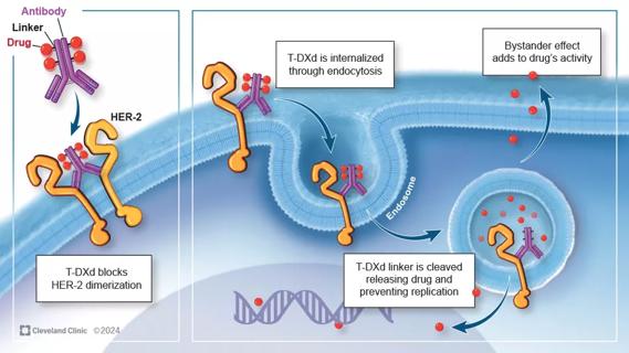
Key learnings from DESTINY trials

Overall survival in patients treated since 2008 is nearly 20% higher than in earlier patients