Locations:

Advice from a real-world case
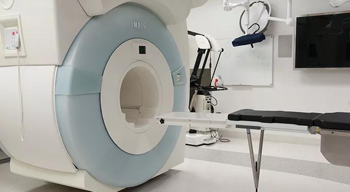
Image content: This image is available to view online.
View image online (https://assets.clevelandclinic.org/transform/795170f8-c865-48d0-817d-bfa3b449785d/Intraoperative-MRI_Joyce_690x380pxl_jpg)
Intraoperative-MRI_Joyce_690x380pxl
Advertisement
Cleveland Clinic is a non-profit academic medical center. Advertising on our site helps support our mission. We do not endorse non-Cleveland Clinic products or services. Policy
By Michael J. Joyce, MD; Nate Mesko, MD; Steven A. Lietman, MD; David Joyce, MD; and Hakan Ilaslan, MD
Musculoskeletal oncologists occasionally encounter tumors that cannot be easily localized during resection, typically because they are not palpable or because of radiation fibrosis or a distorted anatomic field. In such cases, intraoperative MRI— now used by neurosurgeons for intracranial procedures at a number of major medical centers, including Cleveland Clinic— presents an intriguing option. Although the fields yielded by intraoperative MRI are too small for most musculoskeletal cases, they can be adequate for the typical musculoskeletal tumor case, in which MRI localization can provide nearly real time confirmation of tumor margin resection.
We recently reported our first experience with intraoperative MRI-assisted sarcoma resection1 and we recap the case here to showcase the potential benefits of intraoperative MRI in this setting and share insights from our experience.
The case: Myxoid liposarcoma
The patient was a 58-year-old woman with a history of myxoid liposarcoma for which she had had two resections at another institution, each time with positive margins in the proximal right anterior thigh near the femoral vasculature. She later received radiation therapy at another institution.
She then presented at Cleveland Clinic, where we resected satellite recurrences on two occasions nine months apart.
More than a year later, she had local tumor recurrence in two areas of the right thigh. Each was about 2 cm in size and appreciable only on MRI. The surgical field suffered from significant fibrosis and radiation changes, the femoral artery could be localized only with Doppler ultrasonography and the anatomy was markedly distorted. We used intraoperative MRI (principally axial STIR sequences) to confirm the location of both recurrences before incision (Figures 1 and 2).
Advertisement
After incision and approach to the tumor recurrence, the surgical field was scanned to gauge an appropriate margin around each recurrence (Figure 3). After resection, both specimens were placed away from the surgical field but within the intraoperative MRI field to confirm adequate resection and to confirm each specimen with appropriate tissue margins (Figure 4). Postoperative histology confirmed negative margins for both specimens. The patient has remained disease-free over two years of follow-up with serial MRIs.

Image content: This image is available to view online.
View image online (https://assets.clevelandclinic.org/transform/2530edf5-04e0-4b8d-afce-aa0cd4717537/Intraoperative-MRI-Suite_Figs-1-4_Joyce_jpg)
Figures 1 and 2. Intraoperative axial STIR images through the right proximal thigh for localization. The two small, hyperintense soft tissue masses (arrows) are consistent with the known recurrent tumor.
Figure 3. Intraoperative axial STIR image through the right proximal thigh showing surgical flap (arrowheads) elevated over the proximal recurrent soft tissue mass (arrows).
Figure 4. Postoperative axial STIR image showing resected tumor on the patient (arrows at top right) without residual mass in the surgical bed.
What was involved
A neurosurgical MRI suite (Siemens 1.5-tesla magnet on a mobile track) was modified for this case. Using MRI-compatible materials, we worked with hospital engineers to modify the table to allow the patient to be positioned so that the musculoskeletal field (extremity and pelvis) could be visualized with the mobile scanner being moved about the surgical field in a single plane. The cost was $15,000, and changing the table risked voiding its warranty.
Advertisement
The surgical team underwent special training in MRI safety consisting of both an online course and formal magnet room education. Specific protocols were used for surgical instruments and counts to avoid any risk of metal instruments coming under magnetic field influence.
Moving the MRI scanner in and out of the room on tracks took approximately 30 minutes with a 10- to 15-minute scan time. Before surgery, a baseline MRI was obtained for intraoperative comparison. A presurgical “dry run” of the procedure was conducted with the surgical and anesthesiology teams and the operating room nurses.
What we learned
Intraoperative MRI provides a means for addressing local soft tissue sarcoma recurrence in a field with distorted anatomy due to prior resection and changes from radiation therapy. By offering assurance that appropriate margins are being obtained without sacrificing vital structures, intraoperative MRI promises benefits in terms of both patient outcomes and avoidance of repeat surgery.
We conclude that conversion of an MRI surgical suite can be quite helpful for highly select musculoskeletal tumor cases. Planning, flexibility and cooperation among orthopaedic, neurosurgical, radiology and engineering teams are critical. Our advice to other centers considering this is to not underestimate the considerable learning and preparation for MRI suite modification that is required.
Dr. Michael Joyce is Co-Director of the Musculoskeletal Tumor Center. He can be reached at 216.444.4282 or joycem@ccf.org.
Advertisement
Dr. Lietman is Co-Director of the Musculoskeletal Tumor Center. He can be reached at 216.445.2742 or lietmas@ccf.org.
Dr. Ilaslan is a musculoskeletal radiologist in the Department of Diagnostic Radiology. He can be reached at 216.445.4326 or ilaslah@ccf.org.
Dr. Mesko was a resident in the Department of Orthopaedic Surgery when this case was managed. He is now completing a musculoskeletal tumor fellowship and will return to Cleveland Clinic’s Department of Orthopaedic Surgery as a staff physician this fall
Dr. David Joyce was a resident in the Department of Orthopaedic Surgery when this case was managed and is now completing an orthopaedic fellowship
Reference
Advertisement
Advertisement
First-of-its-kind research investigates the viability of standard screening to reduce the burden of late-stage cancer diagnoses
Global R&D efforts expanding first-line and relapse therapy options for patients
Study demonstrates ability to reduce patients’ reliance on phlebotomies to stabilize hematocrit levels
A case study on the value of access to novel therapies through clinical trials
Findings highlight an association between obesity and an increased incidence of moderate-severe disease
Cleveland Clinic Cancer Institute takes multi-faceted approach to increasing clinical trial access 23456
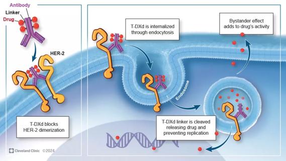
Key learnings from DESTINY trials

Overall survival in patients treated since 2008 is nearly 20% higher than in earlier patients