Locations:

Laser technology and surgery also continue to evolve
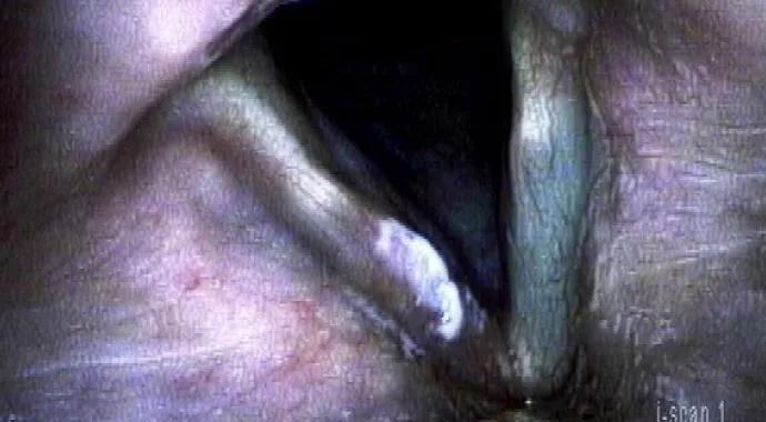
Image content: This image is available to view online.
View image online (https://assets.clevelandclinic.org/transform/1fbd262f-a80b-4e69-976b-18532f023f85/14-ENT-2017-Bryson-Hero-Image-690x380pxl_jpg)
14-ENT-2017-Bryson-Hero-Image-690x380pxl
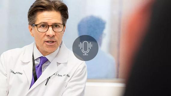
By Paul C. Bryson, MD, and Michael S. Benninger, MD
Advertisement
Cleveland Clinic is a non-profit academic medical center. Advertising on our site helps support our mission. We do not endorse non-Cleveland Clinic products or services. Policy
Laryngeal dysplasia is a premalignant condition of the true vocal folds that is often challenging to treat. Its spectrum of severity ranges from epithelial hyperplasia to severe dysplasia and carcinoma in situ. Complicating the management of laryngeal dysplasia is the variable reliability of tumor grading among pathologists.
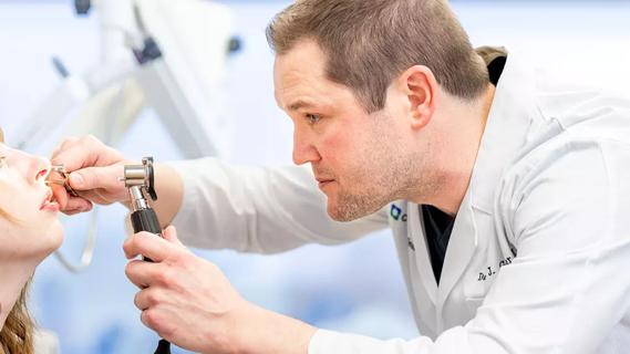
Cleveland Clinic’s Voice Center and Section of Laryngology offer state-of-the-art diagnosis and management of these lesions. Diagnostically, high-definition videostroboscopy (Figure 1), high-speed video and narrow-band imaging (Figure 2) deliver excellent anatomic detail and provide clinicians with the ability to compare lesions over time.
According to traditional teaching on the natural progression of laryngeal dysplasia, it worsens gradually until it becomes a carcinoma. Many clinicians will employ watchful waiting for these lesions, even in patients with a significant voice handicap. The timing and rate of malignant transformation is variable and depends on the severity of disease. Indeed, a large number of patients in Cleveland Clinic’s Head & Neck Institute have what appears to be recurrent but indolent and nonprogressive dysplasia or carcinoma in situ.
Clinically, patients can present with signs and symptoms of varying severity, ranging from minimal voice complaints to severe hoarseness. Endoscopically, laryngeal dysplasia typically appears as patch-like white or red lesions (leukoplakia or erythroplakia) on the surface of the vocal folds. The lesions can dampen mucosal pliability and negatively influence voice. Most patients who develop vocal fold dysplasia have a history of tobacco use, but others have never used tobacco.
Advertisement
The mainstay of treatment has traditionally been microsurgical excision, with or without use of the CO2 laser. Microsurgical excision continues to be the standard with regard to diagnosis, and several advances in laser technology and surgical technique have enhanced our treatment of these lesions in the operating room and the office.
The pulsed potassium titanyl phosphate (KTP) laser has improved the treatment of premalignant vocal fold lesions. The light’s 532-nm wavelength is preferentially absorbed by the subepithelial microcirculation, allowing for the lesion and its surrounding blood supply to be photoangiolysed with high precision. The KTP laser’s fiber-based delivery system also allows patients to be treated comfortably and without sedation in an office-based setting (Figure 3).
Given the propensity of dysplastic lesions to recur, this is a reasonable treatment option for most patients once their disease has been characterized and staged via microsurgical excision, and it may allow them to avoid recurrent general anesthesia. It also allows the laryngeal surgeon to actively treat premalignant disease rather than wait for malignant degeneration.
Malignant transformation is more common in severe dysplasia and carcinoma in situ than in less serious dysplasias, and it can take many years to occur. Therefore, routine surveillance and treatment is our standard of care at the Voice Center. With our state-of-the-art imaging and laser technology, we are able to closely monitor and treat premalignant disease to maintain optimal voice and to survey for possible development of invasive carcinoma.
Advertisement
Dr. Bryson is Director of the Voice Center and Section Head of Laryngology in the Head & Neck Institute.
Dr. Benninger is Chairman of the Head & Neck Institute.
FIGURE LEGENDS
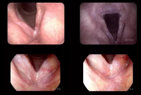
Image content: This image is available to view online.
View image online (https://assets.clevelandclinic.org/transform/d70acf12-16db-4d5d-b4e9-0bc56b41fc7a/14-ENT-2017-Bryson-Inset-Image-01-590pxl-width_jpg)
Figure 1. High-definition videostroboscopic images demonstrate the improved ability to see and characterize the appearance of this lesion on the anterosuperior aspect of the left true vocal fold. The papillary appearance of this lesion is distinct from the surrounding epithelium.
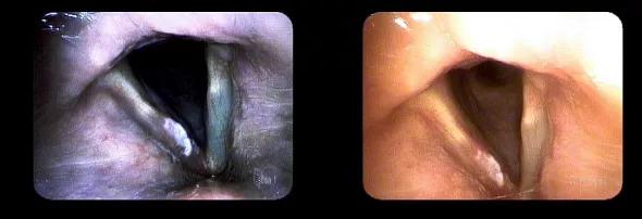
Image content: This image is available to view online.
View image online (https://assets.clevelandclinic.org/transform/02a177f5-6ba4-427e-9147-0e8b17fbab1c/14-ENT-2017-Bryson-Inset-Image-02-590pxl-width_jpg)
Figure 2. These images demonstrate the utility of narrow-band imaging in further clarifying the topographic and angiogenic footprint of a laryngeal neoplasm.
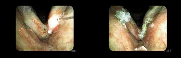
Image content: This image is available to view online.
View image online (https://assets.clevelandclinic.org/transform/a098d778-6657-441c-b457-24bd587283b1/14-ENT-2017-Bryson-Inset-Image-03-590pxl-width_jpg)
Figure 3. Office-based KTP laser photoangiolysis is performed on the severe dysplasia shown in Figure 1. It is performed with the assistance of narrow-band imaging and high-definition endoscopy.
Advertisement
Advertisement

With a wide scope of skills, comprehensive otolaryngologists care for patients of all ages in the community
Research on children with UHL explores the quality-of-life benefits and outcomes of cochlear implants
A look at how custom-fitted oral appliances work and when they’re a good fit for patients
Subtle information gleaned from clinical examinations prompted concern
A new single-port system well-suited for oropharyngeal cancer treatment
Challenging case requires outside-the-box approach
Collaborative and multidisciplinary approach necessary for treatment
The tri-vector gracilis procedure uses a thin muscle from the thigh to help create a natural mimetic smile