Locations:

A case involving cytology and concurrent biopsy
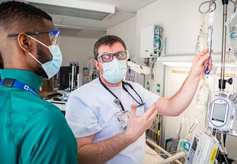
Cytopathologist Charles Sturgis, MD, and thoracic pathologist Sanjay Mukhopadhyay, MD, discuss their diagnostic approach on a case involving pleural fluid cytology and a concurrent pleural biopsy. Watch as they seek answers for an elderly man with a unilateral pleural effusion.
Advertisement
Cleveland Clinic is a non-profit academic medical center. Advertising on our site helps support our mission. We do not endorse non-Cleveland Clinic products or services. Policy
Video content: This video is available to watch online.
View video online (https://www.youtube.com/embed/mcbeU0Gisus?feature=oembed)
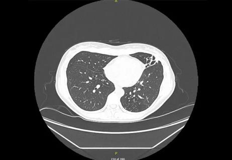
The pathologists begin the discussion with the chest radiograph and CT scan demonstrating the unilateral effusion from the mid-lung field all the way to the base on the right. Dr. Sturgis notes that it is important to learn from the pathologists whether a parenchymal lung lesion is present but that it can be difficult to see anything in the lung parenchyma in large unilateral effusions.
They then examine both the cytology and the histology. Cleveland Clinic Laboratories processes its serous effusion cytologies as two slides, both a ThinPrep® and a cell block. First they discuss a low-power image to emphasize the low overall cellularity of the specimen. On higher magnification, Dr. Sturgis discusses his technique of identifying and comparing a nearby benign cell to the aggregate in question.
In general, adenocarcinomas or metastatic adenocarcinomas appear to have a common border in the group, meaning no extruding nuclei or portions of cells pushing out of the group. In the image in question, the pathologists point out a bit of “hobnailing” where one nucleus extends from the edge of the group, but the majority of the group has more of a common border, with the nuclei within having an increased nuclear to cytoplasmic ratio. Watch the video to learn what type of malignancy the pathologists discover in the pleural fluid.
Cleveland Clinic Laboratories’ Pathology Insights video series features important cases, methods, and practices that are personally presented by our staff pathologists.
Advertisement
These short videos break down information about interesting pathology cases to better inform doctors, laboratory staff, patients or anyone interested in the field of pathology.
Advertisement
Advertisement


Lorem ipsum dolor sit amet. Ut laudantium quasi et aliquam magni qui adipisci sequi et voluptatibus blanditiis et consectetur atque eos nihil dolorem qui culpa nobis. In accusantium voluptatem est modi rerum ut aperiam neque. Hic quos nisi 33 rerum laboriosam cum fuga voluptates. Sed expedita ratione sit nostrum necessitatibus ea maxime perspiciatis sed quia sequi.

This is a subtitle for CQD Post 'Corey Test CQD Post Feb12'. This is a subtitle for CQD Post 'Corey Test CQD Post Feb12'.</end Subtitle>

Matching Secondary -> Primary Snomed Term with Curated Post. Please do not touch this post or any of its categories/snomed terms, thank you!
Lingulectomy removes infection when antibiotics fail
Researchers have developed immunoprofiles for an emerging disease with a mortality rate as high as 27%