Locations:

Radiological findings of different vasculitides
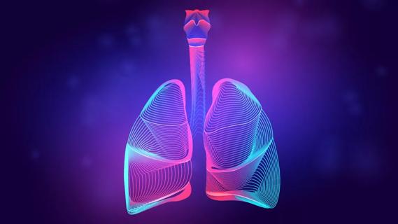
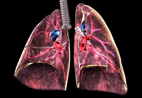
The diagnosis and management of the primary vasculitides is often difficult given that the clinical features of each disease depend on site, degree of severity and other factors, and that many patients present with nonspecific symptoms. Pulmonary vasculitis describes vasculitides with increased involvement of the respiratory system and is usually a facet of systemic vasculitis.
Advertisement
Cleveland Clinic is a non-profit academic medical center. Advertising on our site helps support our mission. We do not endorse non-Cleveland Clinic products or services. Policy
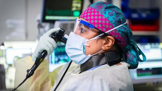
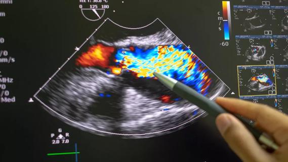
Imaging techniques such as chest radiography, CT and MRI can often help a multidisciplinary team of experts move toward the correct diagnosis. When correlated with laboratory findings, rheumatology and pulmonology consults, imaging can be a vital tool in the diagnosis of pulmonary vasculitis.
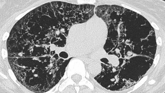
The following images demonstrate the radiologic findings and diagnostic clues of several types of pulmonary vasculitis.

Image content: This image is available to view online.
View image online (https://assets.clevelandclinic.org/transform/d9391193-4d1f-449f-b86a-80d737026f2f/17-RHE-1062-Mahmoud-Slide1-150x103_jpg)
Advertisement

Image content: This image is available to view online.
View image online (https://assets.clevelandclinic.org/transform/9a438056-b57e-477e-aa7b-1644e558bdde/17-RHE-1062-Mahmoud-Slide2-150x103_jpg)

Image content: This image is available to view online.
View image online (https://assets.clevelandclinic.org/transform/901c6703-27fc-4a46-9486-5b12b81407c8/17-RHE-1062-Mahmoud-Slide3-150x103_jpg)
Advertisement

Image content: This image is available to view online.
View image online (https://assets.clevelandclinic.org/transform/ddd7090d-f88e-4f43-b6c6-760acc8b73d1/17-RHE-1062-Mahmoud-Slide4-150x103_jpg)

Image content: This image is available to view online.
View image online (https://assets.clevelandclinic.org/transform/125d8684-10ca-48ea-a411-ed13743f74c7/17-RHE-1062-Mahmoud-Slide5-150x103_jpg)

Image content: This image is available to view online.
View image online (https://assets.clevelandclinic.org/transform/badf204e-1a81-4751-ab08-6393c1c2656d/17-RHE-1062-Mahmoud-Slide6-150x103_jpg)
Slide 1/6
Images and some captions are republished with permission from an article published in Radiologic Clinics of North America. Authors include Shamseldeen Mahmoud, MD, a nuclear radiology fellow; Carol Farver, MD, Director of Pulmonary Pathology; and Joseph Azok, MD, Subha Ghosh, MD, Jason Lempel, MD, and Rahul Renapurkar, MD, all staff in the Imaging Institute.
Advertisement
Advertisement
Advertisement
Volatile organic compounds have potential in heart failure diagnostics
Caregivers are provided with real-time bronchoscopy patient findings
Insights for diagnosing, assessing and treating
A Cleveland Clinic pulmonologist highlights several factors to be aware of when treating patients

New program sets out to better support underserved patient populations
Cleveland Clinic pulmonologists aim to further lower waitlist times and patient mortality
Lessons learned from cohorting patients and standardizing care
New tools and protocols to improve care