Locations:

Cleveland Clinic pediatric spine surgeon shares his insights
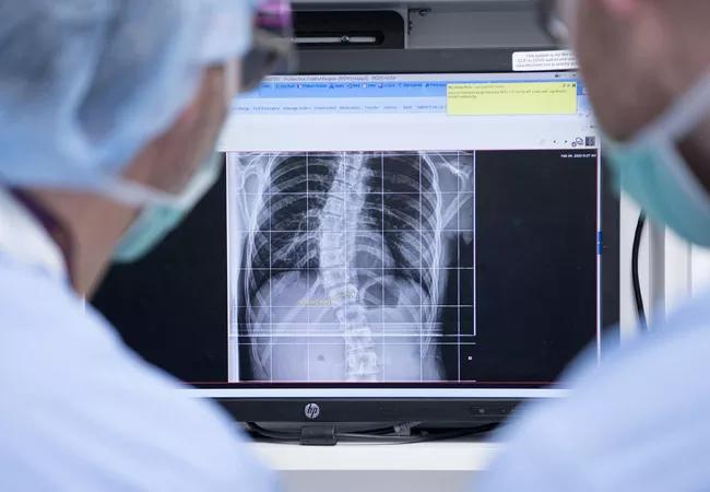
Image content: This image is available to view online.
View image online (https://assets.clevelandclinic.org/transform/01a900c4-5bd5-446f-a9fe-5f39c00bc867/21-ORI-2048688-CQD-Non-fusion-Technique-Scoliosis-2_jpg)
21-ORI-2048688-CQD-Non-fusion-Technique-Scoliosis-2
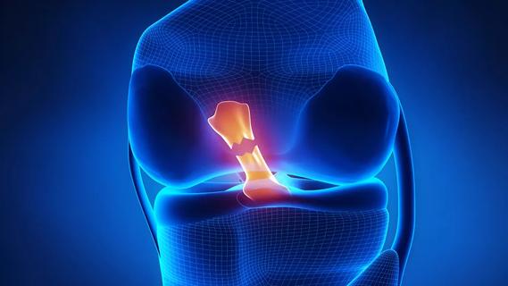
Spinal fusion surgery is the gold standard treatment for severe scoliosis. But it has limitations for young patients who are still growing, as fusion limits their spinal movement and creates additional stress on adjacent segments.
Advertisement
Cleveland Clinic is a non-profit academic medical center. Advertising on our site helps support our mission. We do not endorse non-Cleveland Clinic products or services. Policy
This led pediatric spine surgeon, Ryan Goodwin, MD, to begin incorporating vertebral body tethering (VBT) into his practice. VBT is a minimally invasive technique that modulates continued growth without fusion, preserving motion. Dr. Goodwin, who also directs the Center for Pediatric and Adolescent Orthopaedics in the Department of Orthopaedic Surgery, completed Cleveland Clinic’s first VBT procedure in 2019. He has done over 20 cases to date.
He says he’s heartened by the results he’s seen so far.
Take for example, a case from early 2020 with a 14-year-old patient, an aspiring ballerina. She presented with a curvature that had increased to more than 40 degrees. Dr. Goodwin diagnosed the patient with scoliosis five years prior to this encounter. The curvature, then a moderate 20 degrees, worsened considerably since the initial diagnosis, despite her daily use of a corrective brace for four years.
Dr. Goodwin proposed VBT to the patient and her family. She was a good candidate for the surgery because she was still in an adolescent growth phase, meaning modulation of the curvature – without fusion – was possible.
“There are only a narrow subset of scoliosis patients who are candidates for tethering,” asserts Dr. Goodwin. Pediatric patients with curvatures between 35 degrees and 70 degrees who are refractory to use of a corrective brace may be attractive candidates. Although, as a relatively new procedure, indications continue to evolve.
“Going into the procedure,” he notes, “our goals included stabilization of the convex portion of the curve with non-fusion instrumentation, modest correction in the operating room and completion of the deformity correction with future remaining growth.”
Advertisement
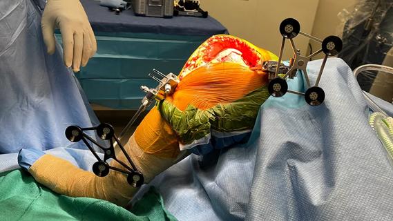
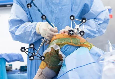
The procedure is performed under general anesthesia in the lateral decubitus position with the convexity up, most typically on the right side. Dual lumen endotracheal tube is preferred so that the lung can be collapsed for visualization. Standard neurologic monitoring is used in all cases. Video-assisted thoracoscopy is performed through four to five portals in the hemithorax.
Video content: This video is available to watch online.
View video online (https://www.youtube.com/embed/--asEvtAvGU?feature=oembed)
Spinal Tethering Surgery
Under camera visualization, as well as fluoroscopic guidance, anterior screws are placed in the vertebral bodies on the convexity along with a staple at each level. This instrumentation is then connected with a tether device that is flexible yet secures the instrumentation under tension, which is controlled by the surgeon.
Image content: This image is available to view online.
View image online (https://assets.clevelandclinic.org/transform/641e10df-a2bc-4ee0-b124-c04ecf31c533/21-ORI-2048688-CQD-Non-fusion-Technique-Scoliosis-8-150x104_jpg)
Image content: This image is available to view online.
View image online (https://assets.clevelandclinic.org/transform/abb6f4f8-b2b3-421b-9d01-1bacb2923e34/21-ORI-2048688-CQD-Non-fusion-Technique-Scoliosis-2-150x104_jpg)
Image content: This image is available to view online.
View image online (https://assets.clevelandclinic.org/transform/9e8817b8-4b3d-4755-89d2-f90788c9b925/21-ORI-2048688-CQD-Non-fusion-Technique-Scoliosis-4-150x104_jpg)
Advertisement
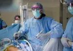
Image content: This image is available to view online.
View image online (https://assets.clevelandclinic.org/transform/7886768a-b123-47f9-a7a0-314cfb47ef45/21-ORI-2048688-CQD-Non-fusion-Technique-Scoliosis-6-150x104_jpg)
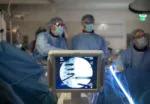
Image content: This image is available to view online.
View image online (https://assets.clevelandclinic.org/transform/a2d0d99f-5868-4cde-a934-b2570748e21f/21-ORI-2048688-CQD-Non-fusion-Technique-Scoliosis-7-150x104_jpg)
Slide 1/5
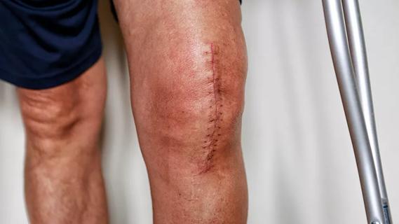
In this particular case, the procedure took four hours. Within a few weeks after the surgery, the patient was back to exercising and taking long walks. Not long after that, she was back to dancing. Dr. Goodwin notes that patients can typically expect to return to full activities without restrictions six weeks postoperatively.
Dr. Goodwin is hopeful that VBT will continue to become a well-utilized technique in the toolkit for scoliosis surgeons, but he cautions, “It’s still relatively new. Over the next 10 to 15 years, it will be important to continue examining the long-term outcomes of this procedure.”
Concluding he notes, “And keep in mind, there is much we still don’t know about the etiology of the disease. In the future, we could potentially be doing even less invasive techniques. But for now, it certainly seems to be a promising approach for treating idiopathic scoliosis in growing children.”
Advertisement
Advertisement

Biologic approaches, growing implants and more
Study reports zero infections in nearly 300 patients
How to diagnose and treat crystalline arthropathy after knee replacement
Study finds that fracture and infection are rare
Center will coordinate, interpret and archive imaging data for all multicenter trials conducted by the foundation’s Osteoarthritis Clinical Trial Network
Reduced narcotic use is the latest on the list of robotic surgery advantages
Cleveland Clinic specialists offer annual refresher on upper extremity fundamentals
Cleveland Clinic orthopaedic surgeons share their best tips, most challenging cases and biggest misperceptions