Locations:

Research points to culprit at the cellular level
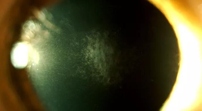
Image content: This image is available to view online.
View image online (https://assets.clevelandclinic.org/transform/83de061d-9648-4745-b396-fb56b8e33766/690x380-Wilson_jpg)
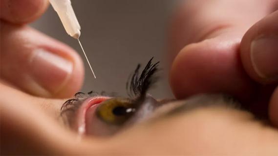
eye corneal scarring
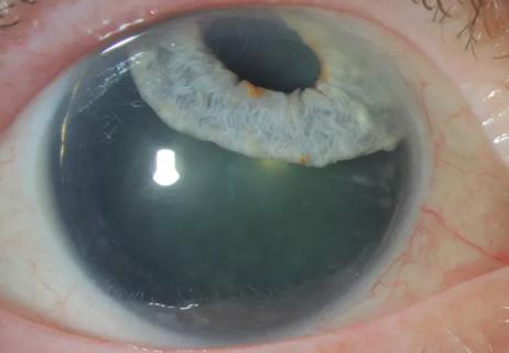
For some people, the search for clear vision free of contact lenses or glasses can lead to an unexpected corneal haze. The culprit, corneal scarring, can be caused by photorefractive keratectomy (PRK) as well as injury, disease or infection, says Steven E. Wilson, MD, Director of the Corneal Wound Healing, Diseases and Ocular Surface laboratory at Cleveland Clinic’s Cole Eye Institute.
Advertisement
Cleveland Clinic is a non-profit academic medical center. Advertising on our site helps support our mission. We do not endorse non-Cleveland Clinic products or services. Policy

Image content: This image is available to view online.
View image online (https://assets.clevelandclinic.org/transform/bd94bc82-4ab2-46bd-845a-c6302e0b49d8/jan2015-subscribe-cqdpulse3_jpg)
One of his lab’s main goals is to identify the molecular and cellular factors that lead to corneal opacity and its resolution. For more than a decade, Dr. Wilson and his colleagues have used innovative rabbit and mouse models to study corneal scarring after high-correction PRK or irregular phototherapeutic keratectomy (PTK), respectively.
Recently, they characterized myofibroblast cells as the main biological factor responsible for the formation of corneal opacity or haze after PRK surgery. In addition, they’ve shown that myofibroblasts come from fibroblastic cells in the cornea (keratocytes) as well as bone marrow-derived cells (fibrocytes) that enter the cornea from the peripheral blood vessels.
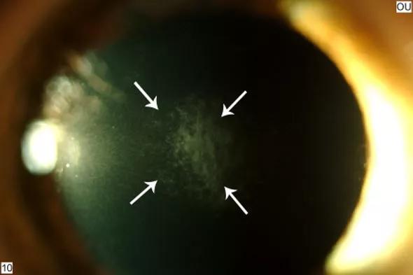
Image content: This image is available to view online.
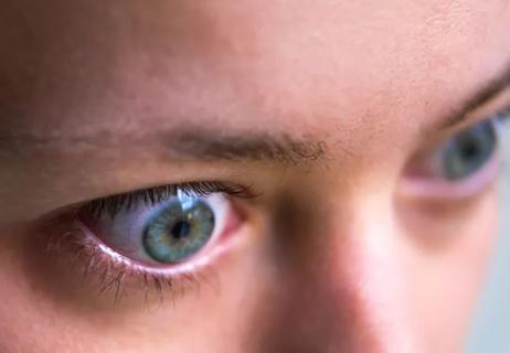
View image online (https://assets.clevelandclinic.org/transform/e215dd25-0bd7-445a-80cc-8f2b1095e175/Fig_-1_-Corneal-haze-3-mo-after-PRK_jpg)
Figure 1. Slit lamp photo of a cornea with haze at 3 months after PRK with myopia. 40X.
After PRK, both kinds of myofibroblast precursor cells begin to differentiate, a process mediated by transforming growth factor beta (TGF-β) and platelet-derived growth factor (PDGF)released by the overlying corneal epithelial cells.
Either type of cell can give rise to myofibroblasts that penetrate the stroma beneath the epithelium and secrete large amounts of disorganized, opaque cellular matrix that’s seen in corneas with late postoperative haze after PRK.
Corneal scarring is also directly related to corneal surface irregularity that can interfere with normal regeneration of the epithelial basement membrane (EBM) after surgery.
During PRK, the central epithelium and the basement membrane are removed. The laser procedure is then performed on the surface of the stroma. When all goes well, the epithelium grows back over the next five to seven days, and the EBM returns to its normal morphology over approximately two weeks.
Advertisement
“In most corneas that undergo PRK,” says Dr. Wilson, “the EBM regenerates completely, epithelium-derived TGF-β and PDGF levels in the stroma dropdue to EBM barrier function, and the myofibroblast precursors stop developing and undergo cell death called apoptosis.”
Recovery goes awry, however, if myofibroblasts develop in the subepithelial stroma and either block production or interfere with localization of one or more epithelial basement membrane component proteins.
These components, which are normally generated by the keratocyte cells in the stroma, need to travel from the stroma to the subepithelial zone where the epithelial basement membrane is being regenerated. However, mature myofibroblasts that may develop in the corneas after high-correction PRK can prevent normal stromal keratocytes from reaching their target site and create a deficiency.
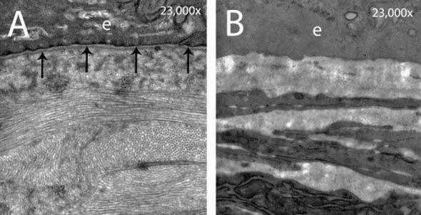
Image content: This image is available to view online.
View image online (https://assets.clevelandclinic.org/transform/ef510997-ebf4-4e66-bc4c-443af4e70057/Fig-2_-A-B_jpg)
Figure 2. Transmission electron microscopy of rabbit cornea A) at 1 month after PRK for –4.5D of myopia and B) 1 month after PRK for –9D of myopia. In clear cornea in A, the lamina lucida and lamina densa (arrows) are visible in the epithelial basement membrane. In cornea with haze in B, no lamina lucida and lamina lucida or lamina densa can be seen. In addition, rows of myofibroblast cells are visible deeper in the central cornea (stroma) but are not seen in clear cornea in A. Magnification 23,000X
“Findings on the critical role of the basement membrane are a major step forward,” says Dr. Wilson. His lab is studying several epithelial basement membrane components, including nidogen-1, nidogen-2 and perlecan. “These are produced by the epithelial cells in the early stages of basement membrane regeneration,” he says, “but after the beginnings of an EBM (nascent EBM) is laid down, they likely must come from the stromal cells such as keratocytes or corneal fibroblasts.”
Advertisement
A common clinically insignificant mild haze occurs in the first few weeks to months after almost all PRK surgeries. However, pathological haze that occurs three to four months post-surgery in humans results from mature myofibroblasts and the excessive disordered extracellular matrix they produce.
“Corneal wound healing following PRK involves a very complex, and at times, unpredictable biological response,” says Dr. Wilson. “A better understanding of cells and molecules involved in the process may lead to new treatment options to restore corneal transparency and prevent corneal scar formation not only after PRK surgery, but also after trauma, infections and in diseases.”
Check out more Cornea articles.
Advertisement
Advertisement
Early data shows risk is 73% higher in patients with lupus, 40% higher in patients with rheumatoid arthritis

Identifies weak spots in the cornea before shape change occurs
Study highlights the value of quantitative ultra-widefield angiography
Switching medications may decrease treatment burden and macular fluid
Interventions abound for active and stable phases of TED
Corneal imaging and interpretation play a major role
Cole Eye Institute imaging specialists are equal parts technician, artist and diagnostician

Effect of low-dose atropine and dual-focus contact lenses is unknown in patients with comorbid eye conditions