Locations:

A glimpse into one of the largest collections of patients imaged at 7T
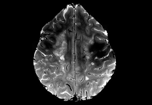
Image content: This image is available to view online.
View image online (https://assets.clevelandclinic.org/transform/7bab83df-be8f-4958-a8ab-17656bdfbc80/16-NEU-568-Jones-650x450_jpg)
16-NEU-568-Jones-650×450
Advertisement
Cleveland Clinic is a non-profit academic medical center. Advertising on our site helps support our mission. We do not endorse non-Cleveland Clinic products or services. Policy
Cleveland Clinic is one of about 50 institutions worldwide with a 7-tesla (7T) MRI scanner — and one of a select few that use it in close relation with an active hospital. An IRB-approved protocol is used to image patients with neurological disease for strict comparison of findings at lower magnetic fields versus 7T.
Over the 18-month period ending in December 2015, Cleveland Clinic used 7T to image 77 patients with the following range of diagnoses:
This collective experience of patients imaged at 7T is one of the world’s largest, and it is serving to help investigate the clinical utility of ultra-high-field MRI.
The principal clinical advantage of 7T imaging stems from increased spatial resolution, including both in-plane voxel spacing and slice thickness. Initial experience suggests that although few lesions are seen at 7T that are not visible at a lower magnetic field, those seen at a higher field are seen with higher resolution and greater neuropathology detail — sometimes leading to an altered diagnosis not appreciated at lower fields. For example, 7T is superior for visualization of microhemorrhages, and it can reveal an increased extent of traumatic brain injury than is seen at lower fields.
Another example is provided by the images below, which show the white matter in a patient with advanced MS. The lower-field (1.5T) image at the bottom shows a uniform field of signal hyperintensity (brightness), but the 7T image at the top allows details within the disease to be appreciated with greater resolution. Note the myriad rounded lesions that appear superimposed at 7T but appear coalesced at lower field.
Advertisement
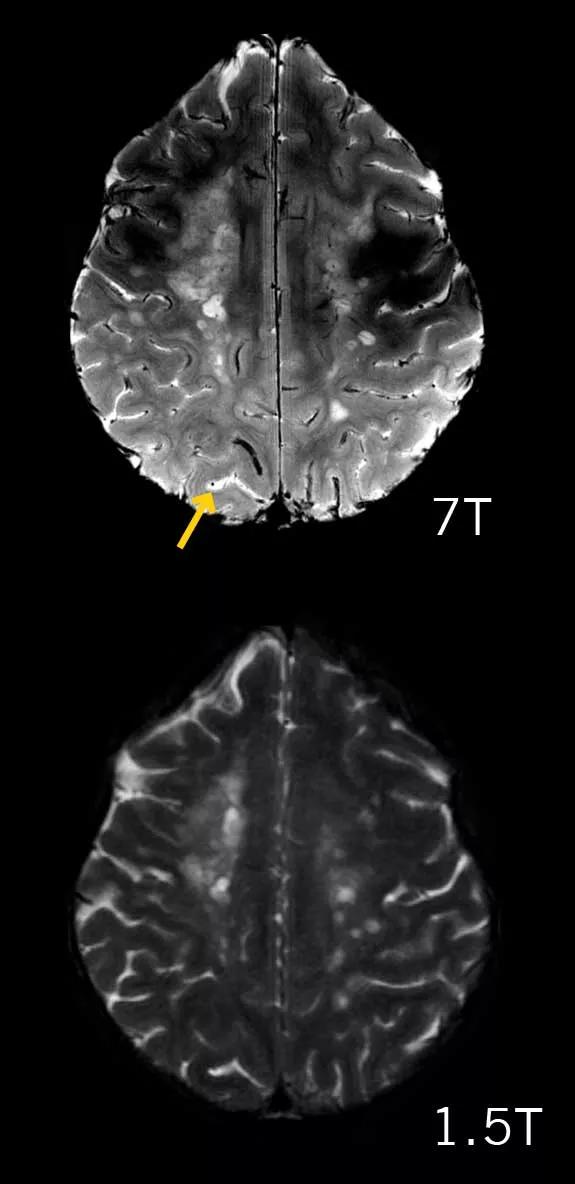
Image content: This image is available to view online.
View image online (https://assets.clevelandclinic.org/transform/d1210f78-c215-46a3-94e6-ee1c6d6745a4/16-NEU-568-Jones-inset_jpg)
MRIs of white matter in a patient with advanced MS taken at 7T (top) and at 1.5T (bottom).
Moreover, 7T reveals a central black dot at the center of nearly all lesions (arrow in the upper image shows one example), which represents the effects of blood flow in a central vein. This association is well established from microscopic pathology, and it can now be demonstrated on MRI with the advent of 7T. In the future, observation of this feature may help determine an MS diagnosis by distinguishing it from numerous look-alikes at lower magnetic field.
Another fascinating advantage of 7T involves resting-state functional imaging, in which brain networks can be revealed with the patient doing nothing in the scanner for a period of six to 10 minutes. The resting-state technique improves visualization as a result of smaller voxels as well as increased blood-oxygen-level-dependent (BOLD) response that scales favorably with magnetic field strength. Recent research with this technique could enhance targeting of intracranial electrodes for deep brain stimulation, for which identification of networks may be essential.
Dr. Jones is Vice Chair for Research and Academic Affairs in Cleveland Clinic’s Imaging Institute and holds appointments in the Epilepsy Center and Mellen Center for Multiple Sclerosis Treatment and Research within Cleveland Clinic’s Neurological Institute.
Advertisement
Advertisement

New study advances understanding of patient-defined goals

Testing options and therapies are expanding for this poorly understood sleep disorder

Real-world claims data and tissue culture studies set the stage for randomized clinical testing

Digital subtraction angiography remains central to assessment of ‘benign’ PMSAH

Cleveland Clinic neuromuscular specialist shares insights on AI in his field and beyond

Findings challenge dogma that microglia are exclusively destructive regardless of location in brain

Neurology is especially well positioned for opportunities to enhance clinical care and medical training

New review distills insights from studies over the past decade