Locations:

Few centers in the U.S. offer this advanced technology

Image content: This image is available to view online.
View image online (https://assets.clevelandclinic.org/transform/b1d53ddd-c3ae-4985-b317-3c39b683c68c/Image-of-Note-690x380_jpg)
Image-of-Note-690×380
In the hands of an experienced surgical team, the O-arm® Surgical Imaging System (Medtronic) allows for a level of precision and safety in spinal surgery that was not possible in the recent past. Several Cleveland Clinic locations ‒ Cleveland Clinic Florida in Weston as well as Center for Spine Health locations at Cleveland Clinic’s main campus and Hillcrest Hospital in Northeast Ohio ‒ are among the few centers in the United States offering this advanced 3-D imaging technology in the OR.
Advertisement
Cleveland Clinic is a non-profit academic medical center. Advertising on our site helps support our mission. We do not endorse non-Cleveland Clinic products or services. Policy
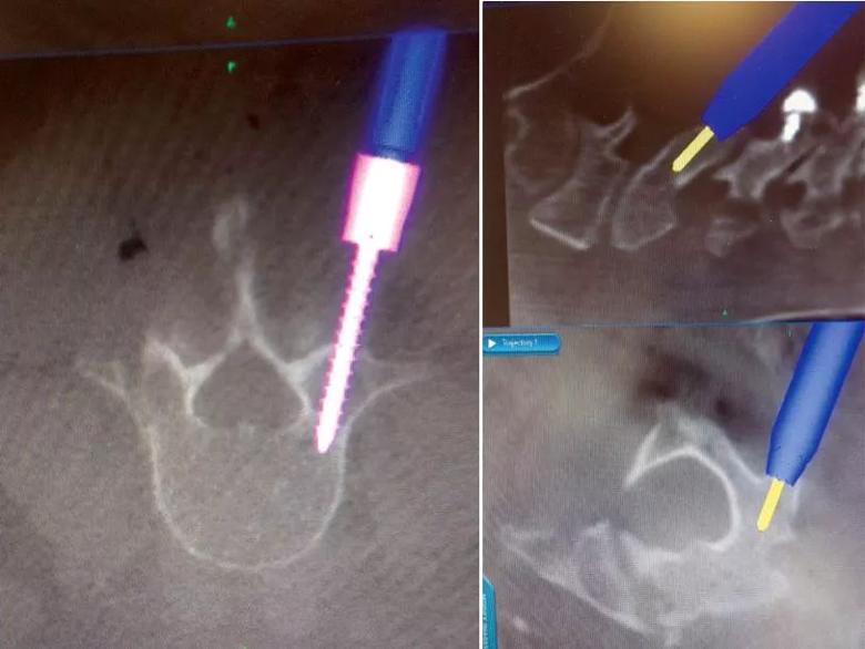
Image content: This image is available to view online.
View image online (https://assets.clevelandclinic.org/transform/b3928cbb-5734-4a80-a35f-d654f05ddbaf/Neuro-post_jpg)
Left: Intraoperative image showing placement of screws in the lumbar spine. Right: Intraoperative images showing placement of screws in the and cervical spine.
In the hands of an experienced surgical team, the O-arm® Surgical Imaging System (Medtronic) allows for a level of precision and safety in spinal surgery that was not possible in the recent past. Several Cleveland Clinic locations ‒ Cleveland Clinic Florida in Weston as well as Center for Spine Health locations at Cleveland Clinic’s main campus and Hillcrest Hospital in Northeast Ohio ‒ are among the few centers in the United States offering this advanced 3-D imaging technology in the OR.
The O-arm is a mobile X-ray system that provides 3-D imaging as well as 2-D fluoroscopic imaging optimized for use in spine surgery. It allows the surgeon to acquire real-time, CT-quality images in the OR that are used to create a 3-D image of the spine. Through integration with the related StealthStation® surgical navigation system, the 3-D image enables superior spinal navigation to facilitate placement of spinal instrumentation with near-perfect accuracy.
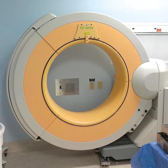
Image content: This image is available to view online.
View image online (https://assets.clevelandclinic.org/transform/03ebb1c2-cdda-4343-9894-54cf9997dc4f/Spine-Image-C_jpg)
Figure. Intraoperative images taken with 3-D computer-assisted surgery enabled by the O-arm Surgical Imaging System.
The O-arm can be used to aid surgery in any portion of the cervical, thoracic or lumbar spine. Its primary uses are for surgical correction of spinal stenosis, spondylolisthesis and spinal deformities. It also can provide 3-D computer-assisted guidance for spinal reconstruction after decompression.
Advertisement
By enhancing visualization of the spinal anatomy, 3-D computer-assisted spinal surgery with the O-arm can significantly improve the safety of spinal procedures and potentially reduce the need for revision surgeries. And because it enables less-invasive approaches, patients stand to benefit from a shortened postoperative recovery period.
Advertisement
Advertisement

New study advances understanding of patient-defined goals

Testing options and therapies are expanding for this poorly understood sleep disorder

Real-world claims data and tissue culture studies set the stage for randomized clinical testing

Digital subtraction angiography remains central to assessment of ‘benign’ PMSAH

Cleveland Clinic neuromuscular specialist shares insights on AI in his field and beyond

Findings challenge dogma that microglia are exclusively destructive regardless of location in brain

Neurology is especially well positioned for opportunities to enhance clinical care and medical training

New review distills insights from studies over the past decade