Locations:

A 68-year-old man with a history of CABG complicated by sternal infection requiring omental and pectoralis flaps presented with a recurrent subxiphoid hernia. Even after three previous repairs with both AlloDerm™ and implanted expanded polytetrafluoroethylene mesh, the sternal defect measured 25 cm wide by 30 cm long. Intestinal contents were herniating from beneath the subxiphoid region and lying on top of the sternum.
Advertisement
Cleveland Clinic is a non-profit academic medical center. Advertising on our site helps support our mission. We do not endorse non-Cleveland Clinic products or services. Policy
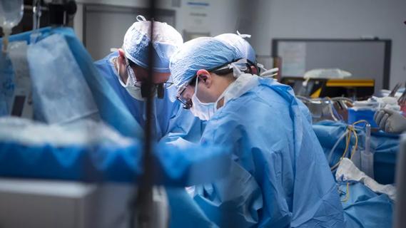
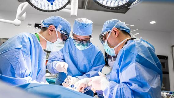
In this brief intraoperative video, watch surgeon Michael J. Rosen, MD, Director of the Comprehensive Hernia Center at Cleveland Clinic, and his team perform the complex reconstruction.
Video content: This video is available to watch online.
View video online (https://www.youtube.com/embed/Zuua6tG_sWs?feature=oembed)
Repair of a Recurrent Subxiphoid Hernia
Dr. Rosen takes special care not to inadvertently enter the hernia sac and injury anything underlying. The team then opens the hernia sac to reveal that both large and small intestine have herniated. The team carefully inspects the bowel to ensure no inadvertent serosal tears are left unrepaired. The surgeons expose the abdominal wall and create a retromuscular space by sizing the posterior rectus sheath approximately half a centimeter from the linea alba. They then carry the incision inferiorly to the pubis taking great care to avoid the inferior epigastric vessels. The lateral dissection is carried out to the linea semilunaris, and further dissection is performed superiorly to the point of the level of the costal margin, a landmark for the beginning of the transversus abdominis release.
Just anterior to the first palpable rib, the posterior lamella of the internal oblique is incised to reveal the transversus abdominis muscle underneath. The surgeons identify the neurovascular bundles and use careful traction and countertraction to discern the planes of the muscles. The posterior lamella of the internal oblique is again incised.
Due to the superior extent of this hernia defect and the need for mesh overlap, Dr. Rosen creates a deep subxiphoid pocket by separating the peritoneum off the overlying diaphragm. He creates a lateral pocket using good traction-countertraction technique. Watch the video to discover what happens once the transversus abdominis has been fully released.
Advertisement
Advertisement

Strong patient communication can help clinicians choose the best treatment option
ctDNA should be incorporated into care to help stratify risk pre-operatively and for post-operative surveillance

The importance of raising awareness and taking steps to mitigate these occurrences
New research indicates feasibility and helps identify which patients could benefit

Treating a patient after a complicated hernia repair led to surgical complications and chronic pain
Standardized and collaborative care improves liver transplantations
Fewer incisions and more control for surgeons

Caregiver collaboration and patient education remain critical