Locations:

The Pediatric Heart Network’s Normal ECG Project
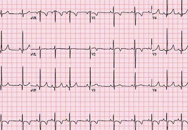
Image content: This image is available to view online.
View image online (https://assets.clevelandclinic.org/transform/035ce1e7-3995-4d43-9385-fe163934baf0/650x450-Saarell_jpg)
650×450-Saarell
Advertisement
Cleveland Clinic is a non-profit academic medical center. Advertising on our site helps support our mission. We do not endorse non-Cleveland Clinic products or services. Policy
The sudden unexplained death of a young person is rare but catastrophic, its impact on families and society incalculable. While prevention of sudden death in the young may be an achievable challenge, many deaths occur without prior symptoms or a family history of sudden death (or at least without recognition of these and their potential connection to sudden death).
The electrocardiogram (ECG), a cornerstone in the evaluation of children with acquired and congenital heart diseases, has the potential to be a useful tool and the basis of screening to prevent sudden cardiac death in the young. However, realizing that potential will require more reliable reference values for pediatric ECGs and a comprehensively better understanding of the role of the ECG in this setting. The National Institutes of Health-sponsored Pediatric Heart Network (PHN) is focusing closely on these goals through its Normal ECG Project, for which I am privileged to serve as the primary investigator. This article reviews the rationale for this project and the insights expected from it.
As a diagnostic tool, the ECG has multiple advantages:
At the same time, the current value of ECGs in the identification and monitoring of heart disease in children is limited by the low frequency of disease and a lack of reliable reference values. The amplitude and duration of surface electrocardiography waveforms are affected by cardiac rhythm, age, gender, heart position and the size of cardiovascular structures; ECG data may also be affected by race and body habitus.
Advertisement
Prior studies defining normal ECG values in children have varied widely in terms of methodology, inclusion criteria, number of subjects and population. Available reports were constructed largely from single sites and have not accounted for analog versus digital ECG recording, geographic variations or the influences of gender and race. Indeed, the most commonly used normal value references in the United States today are derived from analog ECGs acquired in French Canadian children more than 40 years ago.1
The variation in published data and the challenges of using the ECG to identify significant disease provided a strong motivation to obtain more reliable data and create an infrastructure to redefine the modern role of the ECG in the United States and Canada. The PHN, recognizing the dearth of information about normal pediatric ECGs as well as normal pediatric echocardiograms, has undertaken its Normal ECG Project in the hope of ultimately helping prevent sudden death in the young in North America.
This project has widespread implications in that it will obtain an ECG and echocardiogram from a uniformly and accurately defined group of children (all without heart disease) from 19 centers over a broad geographic area in the United States and Canada.
The enrollment target for the PHN Normal ECG Project is 3,600 children from birth to age 18. Findings will be published in 2016.
Significant disagreement exists among experts in the field about the best approach for preventing sudden cardiac death in the young. Some experts support implementation of large-scale cardiovascular screening programs in infants, athletes or all children to identify at-risk individuals in an effort to prevent sudden death. Cardiovascular screening to prevent sudden death typically involves the addition of an ECG to the current standard of care of history-taking and the physical examination. Most who argue against using ECGs for screening in North America cite the lack of reliability of current data to differentiate normal from abnormal children, with unacceptably high false-positive and false-negative rates of detection.
Advertisement
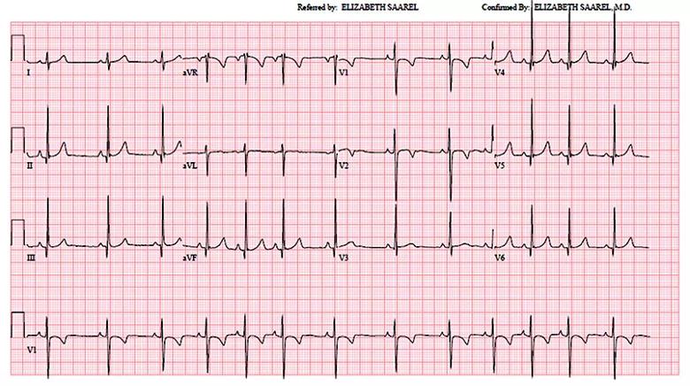
Image content: This image is available to view online.
View image online (https://assets.clevelandclinic.org/transform/d724e002-1f38-401f-a7d1-fde75ba8b6aa/Inset-Saarell_jpg)
Figure. A normal ECG from a 9-year-old boy without heart disease.
The current PHN Normal ECG Project is a retrospective study and thus only a first step on the road to creating an accurate map of normal ECG values for U.S. and Canadian children. There are inherent limitations associated with the quality of the waveforms and with self-reported race in a retrospective investigation. Our hope is that the PHN Normal ECG Project, along with other important studies exploring causes of sudden death in the young, will pave the way for future prospective studies documenting high-quality and accurate standards for ECG values among children in the United States and Canada. Such standards are essential for enabling effective screening to prevent unnecessary and tragic sudden death in the young.
Dr. Saarel, a pediatric electrophysiologist, is Chair of the Department of Pediatric Cardiology. She can be reached at saarele@ccf.org or 216.636.5042.
Advertisement
Advertisement

A sampling of outcome and volume data from our Heart & Vascular Institute

Concomitant AF ablation and LAA occlusion strongly endorsed during elective heart surgery

Large retrospective study supports its addition to BAV repair toolbox at expert centers

Young age, solid tumor, high uptake on PET and KRAS mutation signal risk, suggest need for lobectomy

Surprise findings argue for caution about testosterone use in men at risk for fracture

Residual AR related to severe preoperative AR increases risk of progression, need for reoperation

Findings support emphasis on markers of frailty related to, but not dependent on, age

Provides option for patients previously deemed anatomically unsuitable