Locations:

Two unusual sarcoma cases where necrosis develops in cortical versus medullary bone
By Hakan Ilaslan, MD; Jean Schils, MD; Michael Joyce, MD; Chirag Shah, MD; and Yaxia Zhang, MD
Advertisement
Cleveland Clinic is a non-profit academic medical center. Advertising on our site helps support our mission. We do not endorse non-Cleveland Clinic products or services. Policy
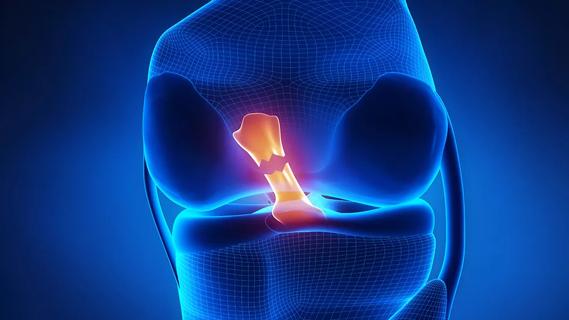
Soft tissue sarcomas often require radiation prior to and after limb-sparing surgery. Post-treatment radiation-induced necrosis is a well-known and potentially severe dose-related complication that usually involves medullary bone. Cortical bone, on the other hand, is considered one of the most radioresistant structures of the body.
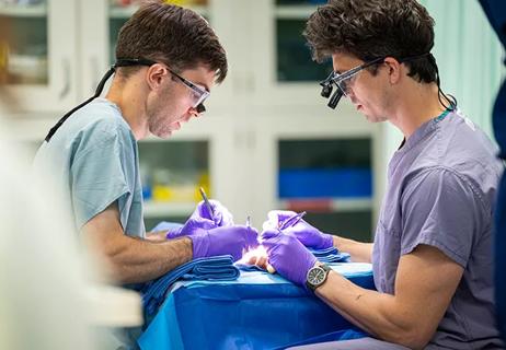
This slideshow features two unusual cases from Cleveland Clinic’s Sarcoma Program/Musculoskeletal Tumor Center. These patients developed radiation-induced necrosis of cortical bone after treatment with neoadjuvant radiation (66 Gy) and surgical resection for soft tissue sarcoma in the thigh. Biopsy of their lesions confirmed isolated circumscribed intracortical necrosis attributed to radiation.
To our knowledge, these are the first reported cases of isolated radiation necrosis of cortical bone.
When bone metastases or recurrent tumor occur after sarcoma treatment, the appearance is typically very aggressive with lytic-destructive behavior, and soft tissue extension is common. Radiation-induced sarcoma may present as a lytic lesion, though it usually appears as very aggressive with cortical destruction and a soft tissue mass. It develops with a median latency period of 12 years after the conclusion of radiation treatment. In the setting of previous high-dose radiation, a well-defined lytic lesion limited to the cortical bone without periostitis may be an early manifestation of radiation necrosis.
Full discussion of these cases can be found in Skeletal Radiology, where they were first reported.
Advertisement

Image content: This image is available to view online.
View image online (https://assets.clevelandclinic.org/transform/7110d725-6d0d-44cf-b1e8-5393784a3646/17-ORT-1274_1-127x150_gif)
Image content: This image is available to view online.
View image online (https://assets.clevelandclinic.org/transform/4a108b09-3fa8-45be-8fd8-208282be0bf2/17-ORT-1274_2-121x150_gif)

Image content: This image is available to view online.
View image online (https://assets.clevelandclinic.org/transform/fcc6a554-ed35-448d-aa1d-65b783efaf9b/17-ORT-1274_3-150x104_gif)
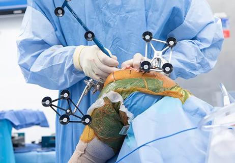
<p>CT-guided biopsy of the lesion with an 11-gauge biopsy needle.</p>

Image content: This image is available to view online.
View image online (https://assets.clevelandclinic.org/transform/f4f4c816-1b4b-4a22-81f4-dc4048545a93/17-ORT-1274_4-150x129_gif)
Advertisement

Image content: This image is available to view online.
View image online (https://assets.clevelandclinic.org/transform/f442b082-6eb7-4853-8313-62497a84fd73/17-ORT-1274_9-129x150_gif)

Image content: This image is available to view online.
View image online (https://assets.clevelandclinic.org/transform/40007207-e114-45e4-ab29-d2165750de3c/17-ORT-1274_6-150x113_gif)
Advertisement
Image content: This image is available to view online.
View image online (https://assets.clevelandclinic.org/transform/41a54d21-acdf-488c-b723-87fb61c95b74/17-ORT-1274_7-7-91x150_gif)

Image content: This image is available to view online.
View image online (https://assets.clevelandclinic.org/transform/5956d35c-a16b-490d-ba80-694ca778382f/17-ORT-1274_8a-84x150_png)

Image content: This image is available to view online.
View image online (https://assets.clevelandclinic.org/transform/2692aee7-3756-4500-81eb-b4e21c9f7f03/17-ORT-1274_8b-100x150_gif)

Image content: This image is available to view online.
View image online (https://assets.clevelandclinic.org/transform/5f166637-68bf-4534-98e7-362a3b1d9286/17-ORT-1274_5-146x150_gif)
Slide 1/10
Dr. Ilaslan is a musculoskeletal radiologist who specializes in bone and soft tissue tumors, and tumor ablations. Dr. Schils is a musculoskeletal radiologist. Dr. Joyce is Co-Director, Musculoskeletal Tumor Center. Dr. Shah is Director of Clinical Research, Department of Radiation Oncology, and Dr. Zhang is a pathologist specializing in bone tumors now at Hospital for Special Surgery.
Advertisement
Advertisement

Biologic approaches, growing implants and more
Study reports zero infections in nearly 300 patients
How to diagnose and treat crystalline arthropathy after knee replacement
Study finds that fracture and infection are rare
Center will coordinate, interpret and archive imaging data for all multicenter trials conducted by the foundation’s Osteoarthritis Clinical Trial Network

Reduced narcotic use is the latest on the list of robotic surgery advantages
Cleveland Clinic specialists offer annual refresher on upper extremity fundamentals
Cleveland Clinic orthopaedic surgeons share their best tips, most challenging cases and biggest misperceptions