Locations:

Exposure can be lowered without affecting test results
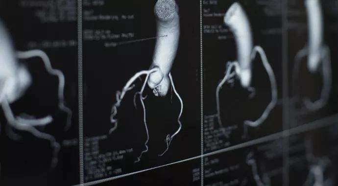
Image content: This image is available to view online.
View image online (https://assets.clevelandclinic.org/transform/5d4c29c3-82bf-464c-ab8c-e90729aeccc9/Radiation-690x380_jpg)
Radiation-690×380
By Samir Kapadia, MD, Director, Cardiac Catheterization Laboratory
Advertisement
Cleveland Clinic is a non-profit academic medical center. Advertising on our site helps support our mission. We do not endorse non-Cleveland Clinic products or services. Policy
With today’s excellent imaging modalities, it’s not uncommon for many patients to have multiple tests for one or more medical problems. For example, a 75-year-old with heart disease may have undergone several stress tests, had two or three cardiac catheterizations and a percutaneous coronary intervention (PCI), a computed tomography (CT) scan performed in the emergency department after a fall, two CTs before and after a hip replacement, and multiple chest X-rays, dental X-rays and mammograms over years of wellness exams.
Although such tests and procedures enable physicians to identify risk and intervene to prevent serious health problems, it is easy to forget that radiation is powerful, cumulative, and needs to be taken seriously.
Patients are not the only ones affected, either. Radiation exposure is a problem for physicians and ancillary personnel, as well as for patients. As of 2012, nine cases of left-sided brain/head-and-neck tumors in interventional cardiologists had been reported. This appears to confirm small epidemiological studies that showed an increased risk of brain tumors in doctors who use fluoroscopy. Radiation exposure may also alter our DNA over time, as studies have shown an increase in chromosomal abnormalities in interventionalists, compared with non-interventionalists.
The deterministic effects of radiation are dose-dependent and include erythema, hair loss, dermal atrophy, fibrosis, desquamation, dermal necrosis, cataracts, decrease in red blood cell production and infertility.
Advertisement
However, malignancies can also occur. The likelihood increases with dose, but the severity is generally independent of dose. In addition to a 20 percent lifetime risk of developing fatal cancer, a lifetime exposure of 100mSv—twice the annual limit of 50mSv for radiation workers—increases the risk by 0.4 percent: 1000 mSv, or what an interventionists might accrue over 20 years in the cath lab—increases the risk by 4 percent.
The key principal, and accepted best practice, is “as low as reasonably achievable (ALARA).”
The amount of radiation exposure delivered by a single test can be lowered by reducing the amount of X-ray delivered. For fluoroscopy, a low dose is considered <50mGy/min, a medium dose 50-100mGy/min and a high dose >100mGy/min.
We have worked with X-ray companies to lower dose acquisition and found we were able to decrease radiation 33 percent without compromising patient care.
Radiation can also be reduced by:
Cineangiography poses a greater challenge, since it delivers 10 to 100 times the dose of fluoroscopy. To minimize radiation:
Advertisement
Question every test. Does the patient need this test? Can it be done with less radiation, or is there an alternate, radiation-free test? Check prior radiation exposures, and if the patient has recently been exposed to more than 5Gy of radiation, consider staging the procedure 6-8 weeks to monitor for skin reaction.
Finally, protect yourself. I wear a lead cap to protect my brain. All interventionalists at Cleveland Clinic wear lead eyeglasses in the cath lab 100 percent of the time.
There are some things you can’t control, such as a patient’s obesity or the need for extreme angles. But using these above guidelines will help protect you and your patients from unnecessary radiation exposure, while delivering the quality of testing you expect.
Maximal annual radiation dose limits for physicians
Advertisement
Advertisement

A sampling of outcome and volume data from our Heart & Vascular Institute

Concomitant AF ablation and LAA occlusion strongly endorsed during elective heart surgery

Large retrospective study supports its addition to BAV repair toolbox at expert centers

Young age, solid tumor, high uptake on PET and KRAS mutation signal risk, suggest need for lobectomy

Surprise findings argue for caution about testosterone use in men at risk for fracture

Residual AR related to severe preoperative AR increases risk of progression, need for reoperation

Findings support emphasis on markers of frailty related to, but not dependent on, age

Provides option for patients previously deemed anatomically unsuitable