Locations:
Search IconSearch
January 11, 2018/Pain Management/Innovation

Quadratus Lumborum Block: A New and Evolving Regional Analgesic Technique (Slideshow)

Strong understanding of the abdominal anatomy is key

Ultrasound-guided injection of local anesthetic in the fascial plane offers anesthesiologists a method that is effective and safe with few complications. One relatively new approach in this realm is the quadratus lumborum (QL) block, a variant of the transversus abdominis plane (TAP) block.

Advertisement

Cleveland Clinic is a non-profit academic medical center. Advertising on our site helps support our mission. We do not endorse non-Cleveland Clinic products or services. Policy

Over the past four years, Hesham Elsharkawy, MD, staff in the Department of General Anesthesiology, and his colleagues at Cleveland Clinic have extensively studied the QL block. They have developed a specialized approach to performing it and have successfully lobbied for a change in the procedure’s nomenclature. “The QL block has advantages,” Dr. Elsharkawy says, “because you can tailor it to different areas of the body such as the lower chest wall, the upper abdomen, lower abdomen and even the hip and lower extremities.”

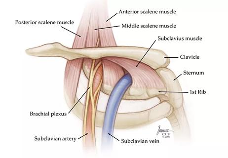
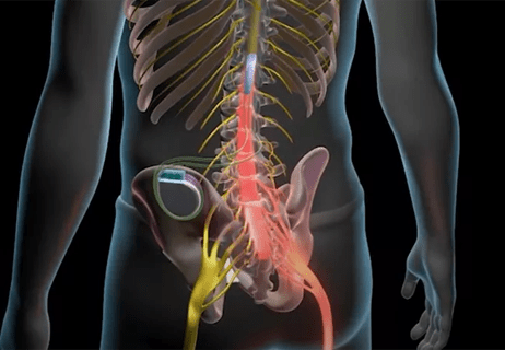
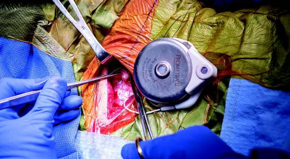
The following illustrations highlight important considerations for physicians performing the QL block. All images are republished with permissions from Elsevier and were originally published in Advances in Anesthesia.

<p>Frontal view of part of the QL muscle with the ventral rami of the spinal nerve roots passing in front of it. The QL muscle is the deep abdominal muscle which stretches from the pelvis to the thorax, allowing it to act as a conduit for local anesthesia between different areas of the body. It lies dorsolateral to the psoas major muscle.</p>

<p>Cross-section view of the anatomy around the QL muscle and posterolateral abdominal wall including the spinal cord, major arteries such as the abdominal branches of the lumbar artery and different fascia including the thoracolumbar fascia (TLF), which is targeted during a QL block. The TLF separates the paraspinal muscles from the QL muscle. It covers the peritoneal surface of the transversus abdominis muscle and continues posteromedially, covering the anterior side of the investing fascia of both the QL and psoas major (PM) muscles. </p>

Advertisement

<p>Two cross-sectional illustrations of the posterolateral abdominal wall with corresponding ultrasound images. The first illustration and ultrasound image show the transverse process view and how it can be used to identify the psoas and the QL muscles. The second show the intertransverse process view and how on ultrasound this view also shows the erector-spinae muscle (ES) and articular process (AP). </p>

<p>Vital structures susceptible to injury during a QL block include the kidney, spleen and liver. The left kidney is slightly higher than the right and ends at the level of the eleventh rib to L2 vertebra, while the right kidney ends at the 12th rib to the upper part of the L3 vertebra. Both are potential injury risks if the block is performed above L3 or if a subcostal approach is used.</p>

<p>Shows the relationship of the kidney with the surrounding fascia. The kidneys are separated from the QL muscle by perinephric fat, the posterior layer of renal fascia and the transversalis fascia. </p>

<p>Researchers believe needle tip placement in relation to the QL muscle is most likely to influence the pattern of local anesthetic spread. Here are three different anatomic locations and the new nomenclature suggested by Dr. Elsharkawy and colleagues.</p><p>1) Lateral: the needle moves from anterior to posterior toward the junction of tapered transversus abdominis muscle and lateral border of QL muscle. It deposits local anesthetic in the lateral border of the QL muscle at the junction with the transversalis fascia and penetrates the aponeurotic attachment of the transversus abdominis muscle.</p><p>2) Posterior: the needle is advanced more posteriorly and deposits local anesthetic posterior to the lateral edge of the QL muscle in the interfascial plane between the posterior border of the QL muscle and the middle thoracolumbar fascia.</p><p>3) Anterior: the needle is advanced either from posterior through erector spinae muscle or anterior through the latissimus dorsi (LD) muscle and then through QL muscle to deposit the local anesthetic in the fascial plane between the QL and psoas major muscles.</p>

Advertisement

<p>These illustrations depict the anterior subcostal paramedian sagittal oblique QL block devel-oped by Dr. Elsharkawy and colleagues. In order to have an oblique sagittal view of the QL muscle, the ultrasound transducer is positioned 6 to 8 cm lateral to the lumbar spinous process at the L1-L2 level with a parasagittal orientation just above the crossover point of the erector spinae and the QL. Then, using a curvilinear transducer with the orientation marker of the ul-trasound cephalad, it is shifted cranially, and the probe is slightly tilted medially. The needle is advanced in plane with ultrasound in a caudal-to-cephalad direction, through the LD then QL muscles. Local anesthetic is deposited anterior to the QL muscle, between the QL muscle and the anterior layer of the thoracolumbar fascia (ATLF). The needle tip is then advanced more cephalad, and local anesthetic, after negative aspiration, is deposited incrementally, observing spread in cephalad direction close to the twelfth rib, with a crescent-shaped distribution of local anesthetic with anterior displacement of the ATLF.</p>

Slide 1/7

Advertisement

Related Articles

20-PAI-1017_CQD_650x450_95843184
February 13, 2020/Pain Management/Innovation
Could Closed-Loop Spinal Cord Stimulation Be the Future of Pain Control?

Randomized controlled study takes a closer look

16-pai-1424-mekhail-hero-image-650x450pxl
September 30, 2016/Pain Management/Innovation
Dorsal Root Ganglion Stimulation Offered for CRPS in Lower Extremities

Experts compare DRG stimulation to traditional SCS

DorsalStim-690&#215;380
October 15, 2015/Pain Management/Innovation
How Dorsal Column Stimulation Helped One Patient Eat Again

Treatment brings near-total relief from abdominal pain

mindfulness
September 21, 2023/Pain Management/Education
Education Program Empowers Patients to Better Manage Chronic Pain

Two-hour training helps patients expand skills that return a sense of control

yoga
Expanding Holistic Pain Management

Program enhances cooperation between traditional and non-pharmacologic care

complex regional pain syndrome
Research Targets Stem Cell Therapy for Complex Regional Pain Syndrome

National Institutes of Health grant supports Cleveland Clinic study of first mechanism-guided therapy for CRPS

thoracic outlet syndrome illustration
Precision Care for Thoracic Outlet Syndrome

Pain specialists can play a role in identifying surgical candidates

Neuromodulation
Therapy Selection in a World of Neuromodulation Options

Individual needs should be matched to technological features

Ad