Locations:

Applications include focal epilepsy, intra- and extracranial tumors
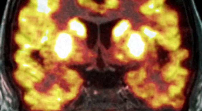
Image content: This image is available to view online.
View image online (https://assets.clevelandclinic.org/transform/e241daf3-f2f5-45da-880e-07583bd5e48f/15-NEU-501-Jones-690x380_jpg)
15-NEU-501-Jones-690×380
Advertisement
Cleveland Clinic is a non-profit academic medical center. Advertising on our site helps support our mission. We do not endorse non-Cleveland Clinic products or services. Policy
During the past decade there has been an imaging revolution in the detection of tumors using combined PET-CT scanners. This union synergistically combines the exquisite sensitivity of fluorodeoxyglucose (FDG) PET for hypermetabolic neoplastic tissue with the high spatial anatomic resolution of CT.
This imaging revolution continues with the recent introduction of combined PET-MRI scanners. Since the spatial resolution of PET is relatively poor compared with MRI, a major advantage of the combination is the ability to use MRI’s superior spatial resolution to accurately identify corresponding values of PET metabolism changes in selected brain subregions. This is particularly useful in pediatric oncology.
Cleveland Clinic recently acquired a leading-edge PET-MRI unit, which resides in the MRI imaging complex on our main campus.
The system utilizes a 3-tesla MRI with a specially developed ring-shaped PET detector inserted in the MRI cowling. The unit is capable of simultaneous scanning, unlike PET-CT, which requires that the imaging acquisitions be obtained sequentially. The time required to obtain a full PET scan easily accommodates a full clinical MRI examination.
Currently our focus is the FDG radiotracer, which is sensitive to tissue metabolic activity; however, in the near future advanced radiotracers will become available.
The image in Figure 1 shows the first pediatric FDG PET-MRI brain examination conducted at Cleveland Clinic, involving a 6-year-old female patient with intractable epilepsy.
Advertisement
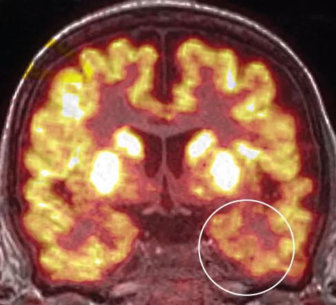
Image content: This image is available to view online.
View image online (https://assets.clevelandclinic.org/transform/f591d494-acd6-4f9e-a6ce-5dc6391b30cd/15-NEU-501-Jones-inset_jpg)
Figure 1. A colorized PET image overlaying a colocalized grayscale anatomic T1-weighted MRI coronal image of a 6-year-old female patient with intractable epilepsy. The white circle is the region of significant hypometabolism involving the left anteromedial temporal lobe and is best appreciated when compared with the same structure on the opposite side.
This coronal imaging shows a colorized PET image overlaying a colocalized grayscale anatomic T1-weighted MRI image. The combined image comes directly from the scanner; that is, the images do not need to be co-registered by hand afterward, as is the current practice, which introduces non-insignificant errors. The PET-MRI image shows significant hypometabolism involving the left anteromedial temporal lobe and reflects the patient’s underlying epileptic disease.
A major benefit of combined PET-MRI scanning for children is the reduced use of sedation. Previously, children who needed both PET and MRI required two separate sedation procedures, each associated with the medical risks of anesthesia and a negative emotional experience. With PET-MRI, both scans can be obtained during a single sedation session. This not only benefits the child and family but is more cost-effective.
The medical benefits of combined PET-MRI scanning for children with neurological disease are multifaceted. As indicated by Figure 1, one advantageous application is for children with focal epilepsy. Children with intracranial or extracranial tumors will also benefit, as will those with inborn errors of metabolism.
Advertisement
Dr. Jones is a neuroradiologist and a staff member of Cleveland Clinic’s Department of Radiology, the Mellen Center for Multiple Sclerosis and the Epilepsy Center.
Advertisement
Advertisement

New study advances understanding of patient-defined goals

Testing options and therapies are expanding for this poorly understood sleep disorder

Real-world claims data and tissue culture studies set the stage for randomized clinical testing

Digital subtraction angiography remains central to assessment of ‘benign’ PMSAH

Cleveland Clinic neuromuscular specialist shares insights on AI in his field and beyond

Findings challenge dogma that microglia are exclusively destructive regardless of location in brain

Neurology is especially well positioned for opportunities to enhance clinical care and medical training

New review distills insights from studies over the past decade