Locations:

Echo is the first, but not always the only, modality
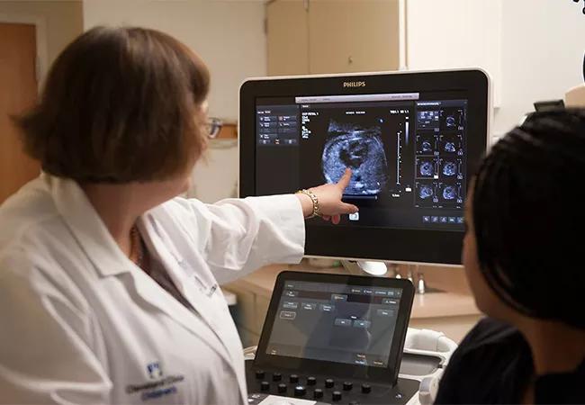
Image content: This image is available to view online.
View image online (https://assets.clevelandclinic.org/transform/98c26deb-c10a-4fdd-8bcd-acee71b52d95/650x450-Erenburg-1_jpg)
650×450-Erenburg
Reasons to refer a baby or child for cardiac imaging can be wide-ranging, running the gamut from cyanosis to syncope to chest pain. Ask Cleveland Clinic Children’s Francine Erenberg, MD, what her go-to imaging test is for diagnosing cardiac issues in babies and children, and there’s no hesitation in her reply.
Advertisement
Cleveland Clinic is a non-profit academic medical center. Advertising on our site helps support our mission. We do not endorse non-Cleveland Clinic products or services. Policy
“Echocardiography is almost always the first test used in diagnosis,” says Dr. Erenberg, a pediatric cardiologist with a specialty interest in echo. Despite that interest, she is quick to add that sometimes MRI or CT is needed to acquire information beyond what echo provides. “For certain patients, echocardiography might not be the only test required.”
Dr. Erenberg notes that echocardiography, or ultrasound of the heart, is the optimal choice for several reasons:
Echocardiography is performed in Cleveland Clinic Children’s echo lab five days a week. In addition to scheduled procedures, the test is available around the clock on a routine and emergent basis for hospitalized children and those needing an immediate diagnosis. “It is especially useful in this regard, because it can be performed at the bedside without moving a critically ill patient,” says Dr. Erenberg.
In obese children, transesophageal echocardiography (TEE) may be needed to obtain a clearer picture. TEE is also routinely used during surgical repair of congenital defects before closure of the chest to make sure the defect has been fixed or the valve works properly.
Most schools that train radiology technicians are oriented toward adult care. So although most hospital radiology departments offer echocardiography, the staff may not be trained to identify CHD in children. This can result in false-negative findings, leaving a child’s diagnosis in limbo.
Advertisement
Cleveland Clinic Children’s echo lab is devoted exclusively to children. “Our technicians receive an additional six to 12 months of training in CHD diagnosis,” says Dr. Erenberg. “Even if the test is negative, we may be able to tell the family what’s wrong with their child.”
Every echocardiogram is performed by a highly trained sonographer and overseen by a pediatric cardiologist with special expertise in imaging. This ensures the study is complete and diagnostic. No sedation or special preparation is required.
A cardiac echo takes 30 to 60 minutes to complete. If a baby or toddler has difficulty lying still long enough to obtain the needed images, the pediatric cardiologist will decide if enough information has been obtained or if the echo must be repeated under sedation.
For all its benefits, echocardiography may not be able to yield complete information in a subset of patients.
MRI may be used as a complementary test when there is suspicion of a problem with the arteries, particularly the pulmonary arteries. MRI also may be used in heavier and more muscular patients, and in those with scarring from previous cardiac surgery. “These conditions can affect the quality of the image taken by the ultrasound transducer,” Dr. Erenberg explains.
MRI and CT can also be used to provide finer detail. “This allows us to assess structures and quantify relationships, such as the size of a defect and the shunt across it, and to obtain information on ventricular and valvular function,” says Cleveland Clinic Children’s Rukmini Komarlu, MD, a pediatric cardiologist with advanced training in cardiac MRI.
Advertisement
The information acquired from MRI or CT can be used to create a three-dimensional model of the heart, which helps with interventions performed in the cardiac catheterization laboratory. “Use of a model like this can shorten the procedure, reducing the child’s radiation exposure,” Dr. Komarlu notes.
Pediatric cardiac CT and MRI are found only in tertiary care centers. “Most hospitals do not have the trained personnel, even if they have scanners for adult patients,” Dr. Komarlu observes.
Cleveland Clinic has three cardiac MRI scanners, including one specifically for pediatrics. Ten to 15 cardiac MRIs are performed daily. Referred patients can obtain a cardiac MRI any day of the week. Pediatric cases that require anesthesia are usually scheduled in advance to ensure availability of a pediatric anesthesiologist. Hospitalized children requiring a cardiac MRI are usually imaged as quickly as possible, depending on scanner availability.
Because abnormal anatomy can be challenging to interpret, all MRI studies on CHD patients are supervised and read by Dr. Komarlu.
MRI scans take 30 to 45 minutes to perform. Patients who cannot remain still require sedation or anesthesia. Teens generally do well if they listen to music or are given antianxiety medication.
Parents may remain in the scanner room and talk to or touch the child during the study. Child life specialists are also present for every procedure in children 12 or younger. They help with the IV line and introduce the child to the scanner. After the study, they award goody bags and take the child’s photo.
Advertisement
CT is a faster study, but because it emits radiation, it is reserved for situations when MRI cannot be performed, such as when the child has a pacemaker, defibrillator, aneurysm clip, coil, bullet or other metal object in the body. A CT may also be ordered to provide information about arterial structure or kidney function.
Referring physicians provide valuable assistance by informing imaging specialists about any metal in the patient’s body before referral for imaging.
Advertisement
Advertisement

A sampling of outcome and volume data from our Heart & Vascular Institute

Concomitant AF ablation and LAA occlusion strongly endorsed during elective heart surgery

Large retrospective study supports its addition to BAV repair toolbox at expert centers

Young age, solid tumor, high uptake on PET and KRAS mutation signal risk, suggest need for lobectomy

Surprise findings argue for caution about testosterone use in men at risk for fracture

Residual AR related to severe preoperative AR increases risk of progression, need for reoperation

Findings support emphasis on markers of frailty related to, but not dependent on, age

Provides option for patients previously deemed anatomically unsuitable