Locations:

Localizing the site of hyperactivity that causes seizures
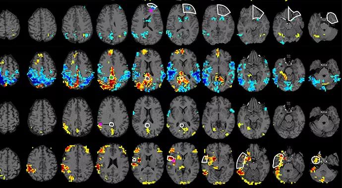
Image content: This image is available to view online.
View image online (https://assets.clevelandclinic.org/transform/d9eb31fd-ded3-4176-975b-7619f097381f/Jones-690x380_jpg)
Jones-690×380
At first blush, the cure for pharmacoresistant focal epilepsies ‒ surgical removal of the brain’s epileptogenic zone (EZ) ‒ can seem deceptively straightforward. The trick, however, is ensuring sufficient identification of the EZ and related epileptic network. Several techniques have been proposed, but none has offered adequate spatial coverage and sufficiently precise localization of source signals. Until now.
Advertisement
Cleveland Clinic is a non-profit academic medical center. Advertising on our site helps support our mission. We do not endorse non-Cleveland Clinic products or services. Policy
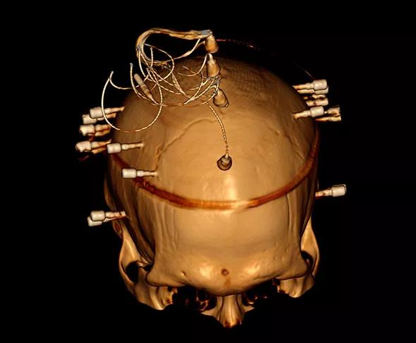
Image content: This image is available to view online.
View image online (https://assets.clevelandclinic.org/transform/4e60ba61-3c96-49f5-8a2a-2c59cda9afd4/Jones-inset2_jpg)
In May 2014, a Cleveland Clinic team led by neuroradiologist Stephen E. Jones, MD, PhD, and neurosurgeon Jorge Gonzalez-Martinez, MD, PhD, published preliminary clinical data (BrainConnect. 2014;4[4]:286-298) introducing a technique for studying brain connectivity that promises to enhance preoperative identification of epileptic areas in the human brain.
Their technique arises from the earlier observation that the blood-oxygen-level-dependent (BOLD) response on fMRI can be used to demonstrate localized networks of activity across the entire brain, usually through task-related activation or while the patient is at rest. Working from this premise, the investigators decided to combine direct intracranial electrical stimulation of the brain with simultaneous fMRI (DES-fMRI) to assess the BOLD response to intracranial stimulation near the hypothesized EZ. They then compared the response to evoked electrical recordings from other intracranial electrodes.
The team reported results from the first five patients with pharmacoresistant seizures who underwent the DES-fMRI technique at adequate stimulation frequencies and voltages. All patients were already undergoing invasive evaluation for presurgical planning using the stereoelectroencephalographic (SEEG) methodology.
Robust fMRI maps of activation networks were produced easily, and they showed a significant but weak positive correlation between quantitative measures of BOLD activity and measures of electrical activity in response to direct stimulation.
Advertisement
Among the four patients with outcomes data at six months after surgery, successful surgical outcome was consistent with the resection of brain regions that had high local fMRI activity.
“We were able to stimulate the electrodes during simultaneous fMRI imaging and see in real time how the entire brain ‒ four-dimensionally ‒ reacted to the stimulation,” says Dr. Jones. “This allows us to localize the site of hyperactivity that causes the patient’s seizures.”
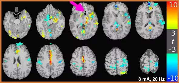
Image content: This image is available to view online.
View image online (https://assets.clevelandclinic.org/transform/395b3c1f-ce20-47ba-a78b-8c8495b717f6/Jones-inset1_jpg)
fMRI activation maps during high-current stimulation trials in a patient. The site of bipolar stimulation is marked by the two asterisks denoted by the large magenta arrow. The overlaid colored regions indicate portions of the brain that significantly react to the stimulation, as denoted by the color bar (red indicates increased reactivity; blue indicates reduced activity). Note how the brain can react to stimulation both close to and far away from the electrodes ‒ even on the opposite side of the brain. Most interestingly, the pattern of activation conforms to segments of known functional anatomy (e.g., the cingulate gyrus).
The results suggest a role for DES-fMRI in evaluation of full-brain volume in studies of brain connectivity, including in surgical planning for patients with medically intractable epilepsy. Since the BOLD response was found to be consistent with electrical recordings, the investigators note that the complete coverage and precise localization offered by fMRI could complement invasive evaluation, extending the range of corticocortical evoked potential assessment to the whole brain.
Advertisement
The technique also might improve the yield of invasive evaluations by identifying brain regions with high activity levels that merit sampling via a SEEG electrode. Such a procedure could be done intraoperatively and interactively, with the DES-fMRI data from each electrode guiding the placement of the subsequent electrode.
“Sometimes we need to perform two or three surgical procedures to control the seizures in a patient with refractory focal epilepsy,” says Dr. Gonzalez-Martinez. “This technique promises to better define the seizure area so we can cure these patients with a single procedure.”
As in most biomedical advancements, the team’s investigation prompted at least as many questions as it answered. Among the questions to be addressed by their ongoing and future studies:
Regardless of exactly what these future investigations yield, Dr. Jones suspects their team has come across an important real-time combination of technologies. “If you can help identify the one piece of tissue implicated in refractory seizures,” he notes, “you can have a tremendous impact on a patient’s life.”
Advertisement
Advertisement

New study advances understanding of patient-defined goals

Testing options and therapies are expanding for this poorly understood sleep disorder

Real-world claims data and tissue culture studies set the stage for randomized clinical testing

Digital subtraction angiography remains central to assessment of ‘benign’ PMSAH

Cleveland Clinic neuromuscular specialist shares insights on AI in his field and beyond

Findings challenge dogma that microglia are exclusively destructive regardless of location in brain

Neurology is especially well positioned for opportunities to enhance clinical care and medical training

New review distills insights from studies over the past decade