Locations:

Valgus tibiotalar tilt tough to treat, common
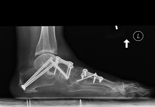
Image content: This image is available to view online.
View image online (https://assets.clevelandclinic.org/transform/f80f67f4-c1e5-48c2-bbc9-cbb1610a452c/16-ORT-2647-Miniaci-ConsultQD_650x450_jpg)
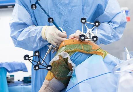
16-ORT-2647 Miniaci ConsultQD_650x450
By Sara Lyn Miniaci-Coxhead, MD
Advertisement
Cleveland Clinic is a non-profit academic medical center. Advertising on our site helps support our mission. We do not endorse non-Cleveland Clinic products or services. Policy
Arthrodesis of the hindfoot is a common, reliable option for treatment of arthritis and deformity involving the subtalar, talonavicular and calcaneocuboid joints. The development of valgus tibiotalar tilt of the ankle joint has been noted as a complication of these procedures. Only limited data examine the incidence and predictive factors of valgus tibiotalar tilt following hindfoot fusion. We sought with this study to define the incidence of valgus tibiotalar tilt, and to identify any pre- or postoperative radiographic parameters that may predict its postop development.
Valgus tibiotalar tilt is a tough phenomenon to treat given deltoid ligament insufficiency and the limited options for deltoid ligament reconstruction. In addition, ankle joint incongruity leads to accelerated wear due to increased eccentric contact pressures. This can lead to ankle arthritis, another difficult problem with limited surgical treatment options.
We retrospectively reviewed all patients over a 10-year period who underwent a subtalar, subtalar and talonavicular (double), or subtalar, talonavicular and calcaneocuboid (triple) arthrodesis. Exclusion criteria included an ankle arthrodesis, a revision procedure, total ankle arthroplasty or a deltoid ligament reconstruction at the time of the index procedure. Patients were also excluded if the indication for surgery was a cavus foot deformity or Charcot arthropathy, or if they did not have a complete set of weight-bearing foot and ankle radiographs (pre- and postoperative).
Advertisement
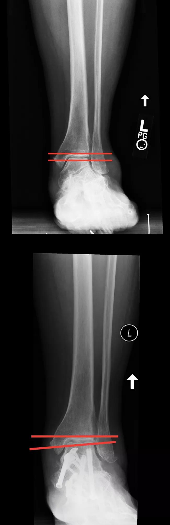
Image content: This image is available to view online.
View image online (https://assets.clevelandclinic.org/transform/d7e2f35c-5450-4d8e-82ff-58590962958d/16-ORT-2647-Miniaci-ConsultQD-Inset-1_jpg)
Figure 1. Preop (above) and postop (below) radiographs of a patient who developed tibiotalar tilt postop.
A total of 187 patients were eligible for the study. The average age was 52 (11 to 82) years. The most common indication for surgery was adult-acquired flatfoot deformity (92, 49.2 percent), followed by arthritis (83, 44.4 percent). The most common procedure was a triple arthrodesis (101, 54 percent). A total of 51 patients (27.3 percent) developed valgus tibiotalar tilt postoperatively, at an average time of 3.6 months after surgery (Figure 1).
Standardized measurements were taken on both pre- and postoperative films. These measurements were previously described as appropriate in the evaluation of pes planus. We found that an increase in preoperative Meary’s (lateral talar-first metatarsal) angle (hazard ratio 1.039, CI: 1.002-1.077; P < 0.05) was associated with development of valgus tibiotalar tilt. An increase in postoperative Meary’s angle (hazard ratio 1.052, CI: 0.999- 1.108, P = 0.0528) approached significance for development of tibiotalar tilt. We found that patients with larger or undercorrected deformities were more likely to develop tibiotalar tilt.
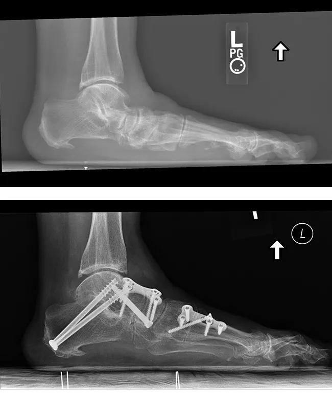
Image content: This image is available to view online.
View image online (https://assets.clevelandclinic.org/transform/ec6463f1-3de0-4a9c-8056-216d76ee5c87/16-ORT-2647-Miniaci-ConsultQD-Inset-2_jpg)
Figure 2. A. A large preoperative deformity. B. An undercorrected deformity.
A review of multiple cadaver studies revealed that the development of valgus tibiotalar tilt appears to be related to strain and incompetence of the deltoid ligament. In a patient with a flatfoot deformity, weight-bearing forces dissipate throughout the hindfoot, allowing the tibiotalar joint to remain in appropriate alignment. However, once the hindfoot is fused, it lacks flexibility. This rigidity, combined with deltoid ligament attenuation, leads to eccentric forces through the tibiotalar joint, producing valgus tibiotalar tilt.
Advertisement
This information has an important impact on our surgical practice. First, high-risk patients should be informed about the risks and consequences of valgus tibiotalar tilt.
Operative planning should center on a reconstruction-based procedure rather than a fusion when feasible, as this would allow hindfoot flexibility. Also, surgeons should assess and consider possible reconstruction of the deltoid ligament at the time of correction, in hopes of preventing tibiotalar tilt.
Our ongoing research in this area will compare the outcomes of patients who do and do not develop tibiotalar tilt. We are also examining the role of deltoid ligament stress during a hindfoot fusion and the efficacy of deltoid ligament reconstruction at the time of the index procedure. The more we learn about the development and impact of tibiotalar tilt, the more we can avoid further morbidity for our patients.
Dr. Miniaci-Coxhead is associate staff in the Department of Orthopaedic Surgery. She would like to acknowledge coauthors for this project, A. Samuel Flemister, MD, and John P. Ketz, MD, at the University of Rochester, Department of Orthopaedics and Rehabilitation, in Rochester, NY.
Advertisement
Advertisement

Biologic approaches, growing implants and more
Study reports zero infections in nearly 300 patients
How to diagnose and treat crystalline arthropathy after knee replacement
Study finds that fracture and infection are rare
Center will coordinate, interpret and archive imaging data for all multicenter trials conducted by the foundation’s Osteoarthritis Clinical Trial Network

Reduced narcotic use is the latest on the list of robotic surgery advantages
Cleveland Clinic specialists offer annual refresher on upper extremity fundamentals
Cleveland Clinic orthopaedic surgeons share their best tips, most challenging cases and biggest misperceptions