Locations:

Cleveland Clinic study demonstrates systems’ advantages
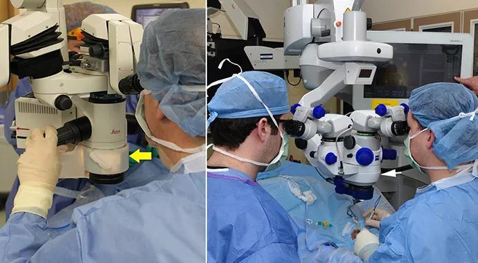
Image content: This image is available to view online.
View image online (https://assets.clevelandclinic.org/transform/d73b6a07-f721-4c6f-a6e6-d567c09ee8f0/690x380-Ehlers-Discover_jpg)
690×380-Ehlers-Discover
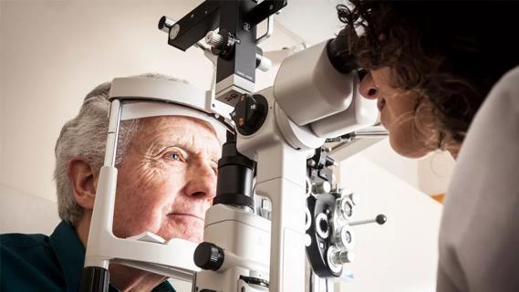
While optical coherence tomography (OCT) provides cross-sectional, near-histology-level detail on tissues of interest in ophthalmic surgery, the complexity of creating a system that allows for minimal intrusion into the operating room and seamlessly integrating into the surgical microscope is a significant challenge, says Justis Ehlers, MD, a staff physician on the vitreoretinal service of Cleveland Clinic’s Cole Eye Institute.
Advertisement
Cleveland Clinic is a non-profit academic medical center. Advertising on our site helps support our mission. We do not endorse non-Cleveland Clinic products or services. Policy

Image content: This image is available to view online.
View image online (https://assets.clevelandclinic.org/transform/bd94bc82-4ab2-46bd-845a-c6302e0b49d8/jan2015-subscribe-cqdpulse3_jpg)
The widespread adoption of OCT has transformed the treatment of ophthalmic diseases. The recent DISCOVER study confirms the feasibility of a further advancement in state-of-the-art ophthalmic surgery. The device study, conducted at Cleveland Clinic Cole Eye Institute, examined the use of the microscope-integrated intraoperative optical coherence tomography (iOCT) systems with surgeon feedback and surgeon control in anterior segment and posterior segment surgery. Various findings from this large-scale study have been published in the British Journal of Ophthalmology, JAMA Ophthalmology and the American Journal of Ophthalmology.
Microscope-integrated iOCT technology allows for rapid visualization of the impact of surgical maneuvers on the tissues of interest, delivering immediate feedback on the completion of surgical objectives (e.g., extent of membrane peel, optimal graft placement).
It also enables surgeons to immediately address areas of need, such as tissue architecture and and microstructure, and feel confident of success in the achievement of surgical goals.
The study found that the iOCT system successfully allowed for outstanding visualization of tissues during surgery without disrupting surgical flow significantly.
“Over the last few years, significant advances have been made on integrative technology, both as add-ons to current microscopes and as complete integrated microscope-OCT systems,” Dr. Ehlers says.
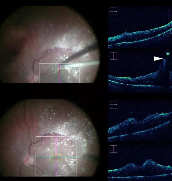
Image content: This image is available to view online.
View image online (https://assets.clevelandclinic.org/transform/147d03e5-1c80-455e-8789-7b773153bb2c/590x-Inset-Ehlers-Discover_jpg)
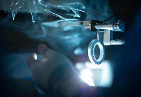
The DISCOVER trial is examining three different microscope integrated OCT systems, including the Zeiss prototype Rescan 700, the Bioptigen integrated prototype system and an internally developed the Cole Eye integrated system. These systems include various display systems (e.g., heads-up, external screen) and surgeon controls (e.g., mouse-based, foot-pedal).
Advertisement
iOCT scanning took place during key milestones with continuous visualization and motion. In addition, the systems enabled iOCT capture of volume and raster scans. Surgeons could customize select the scan length, angle and location.
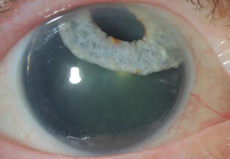
Results showed that iOCT images were successfully obtained in the vast majority of cases. Anterior segment OCT visualization included corneal incisions, corneal graft orientation/position, phacoemulsification groove depth and intraocular lens position. Posterior segment images included evaluation of hyaloid release, completeness of membrane peeling and retinal detachment cases.
The iOCT systems allowed for real-time visualization of tissue-instrument interactions. Metallic instruments resulted in significant shadowing of underlying tissues. Silicone instruments exhibited improved OCT transmission. Optimizing OCT-compatible instrumentation is an ongoing area of development for iOCT.
“The technology is still emerging and at an early stage,” says Dr. Ehlers. He reported that challenges remain in image quality in the surgical environment, OCT-compatible instrumentation, software analysis and automated aiming. “These are all areas of active development at the Cleveland Clinic Cole Eye Institute,” he says.
The U.S. Food and Drug Administration has cleared two microscope-integrated iOCT systems, the Zeiss RESCAN 700 and Haag-Steit iOCT.
“The overall interest in and adoption of intraoperative OCT continues to increase, but continued research is needed to better elucidate the optimal roles for iOCT in enhancing patient outcomes,” he says.
Advertisement
Advertisement
Early data shows risk is 73% higher in patients with lupus, 40% higher in patients with rheumatoid arthritis

Identifies weak spots in the cornea before shape change occurs
Study highlights the value of quantitative ultra-widefield angiography
Switching medications may decrease treatment burden and macular fluid

Interventions abound for active and stable phases of TED
Corneal imaging and interpretation play a major role
Cole Eye Institute imaging specialists are equal parts technician, artist and diagnostician

Effect of low-dose atropine and dual-focus contact lenses is unknown in patients with comorbid eye conditions