Locations:

Shapshots from our applications in MS and epilepsy
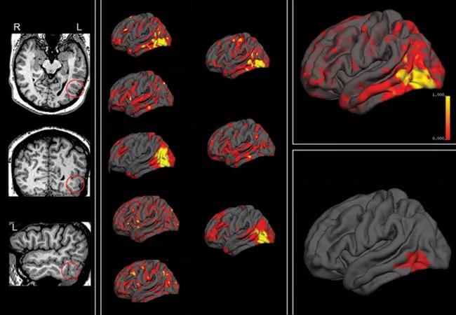
Image content: This image is available to view online.
View image online (https://assets.clevelandclinic.org/transform/02091b3a-c3db-4ca1-b4bf-4476365bd19e/18-NEU-508-Nakamura-MRI-650x450_jpg)
18-NEU-508-Nakamura-MRI-650×450
By Kunio Nakamura, PhD, and Z. Irene Wang, PhD
Advertisement
Cleveland Clinic is a non-profit academic medical center. Advertising on our site helps support our mission. We do not endorse non-Cleveland Clinic products or services. Policy
The advent of artificial intelligence and machine learning is revolutionizing the way large volumes of patient data are interpreted. Scientists in Cleveland Clinic’s Neurological Institute are devoting substantial efforts to using machine-learning algorithms to inform the way pathological substrates are being detected and delineated in imaging studies. This post briefly profiles our work in this area in two major neurological diseases — multiple sclerosis (MS) and epilepsy.
One innovative application of machine learning is for quantification of MS lesion loads from clinical imaging studies. Researchers with Cleveland Clinic’s Lerner Research Institute and Mellen Center for Multiple Sclerosis Treatment and Research have analyzed MRIs acquired on clinical scanners using a Random Forest algorithm, which improves overall segmentation accuracy over other methods when compared with consensus segmentation from multiple reviewers (Figure 1).
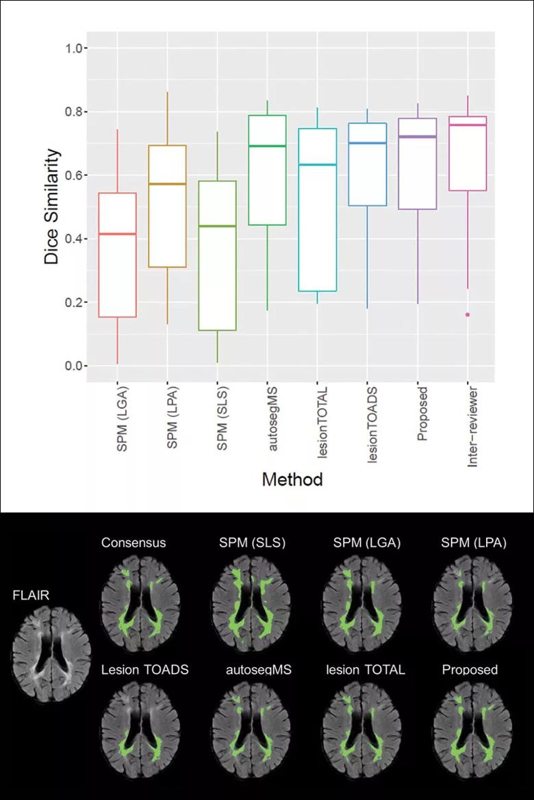
Image content: This image is available to view online.
View image online (https://assets.clevelandclinic.org/transform/c5c1d8c1-a72e-4c58-be54-4be92826374d/18-NEU-508-Nakamura-MRI-inset1_jpg)
Figure 1. Top: Box plots of Dice similarity measures from various MS lesion segmentation methods. The Dice ranges from 0 to 1, with 0 being no overlap and 1 being perfect overlap. Our proposed machine-learning method (second plot from the right) showed the highest mean Dice measure. Although far from achieving perfect overlap, our method is approaching the range of inter-reviewer variability (rightmost plot). Bottom: Example of visual comparison of lesion segmentation methods.
Notably, the algorithm is able to analyze MRIs from various clinical protocols and from different MRI models, including field strengths of 1T, 1.5T and 3T, as well as from various scanner manufacturers (Figure 2).
Advertisement
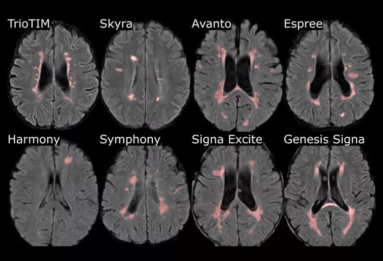
Image content: This image is available to view online.
View image online (https://assets.clevelandclinic.org/transform/ca9f6355-bbc2-469b-a25f-7c5939e0d3ea/18-NEU-508-Nakamura-MRI-inset2_jpg)
Figure 2. T2 lesion segmentation from eight different scanner models showing the applicability in various clinical MS protocols.
When compared with clinical reads by neuroradiologists, the automatic lesion volumes showed good associations between severity categories and visually confirmed the lesion probability maps based on the categories (Figure 3). These findings highlight the usefulness of machine-learning techniques and suggest a potential to change routine practice.
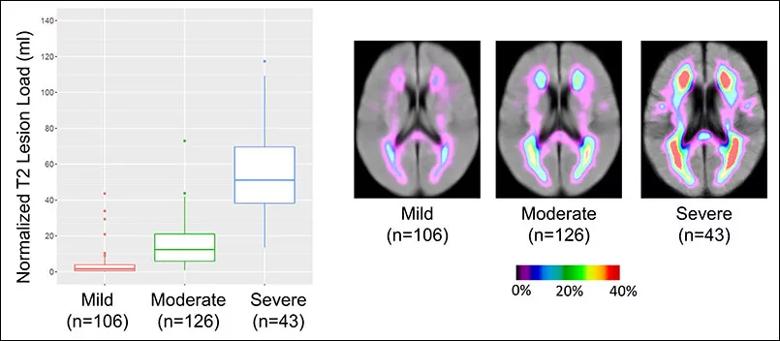
Image content: This image is available to view online.
View image online (https://assets.clevelandclinic.org/transform/bce6471d-3258-4a42-983a-cb61f369663a/18-NEU-508-Nakamura-MRI-inset3_jpg)
Figure 3. Left: Box plots of normalized T2 MS lesion volume according to clinical reads. Right: Average lesion probability map overlaid on average fluid-attenuated inversion recovery (FLAIR) images for each clinical category. The images show increasing lesion probability in periventricular and subcortical areas. Concurrently, an enlargement of the ventricles is seen as clinical severity intensifies from mild to severe.
Equipped with a large surgical volume, MRI data and pathological confirmation of resected tissue, the research group in Cleveland Clinic’s Epilepsy Center is investigating machine-learning strategies designed to directly correlate MRI with pathological findings.
Figure 4 shows an automated multivariate surface-based morphometry analysis methodology combined with machine learning for automated detection of focal cortical dysplasia (FCD). The analysis was based on measures such as cortical thickness, gray-white matter blurring, sulcal depth and curvature, and cortical shape deformation. Additionally, asymmetry maps were generated to account for inherent left-right differences in each individual brain.
Advertisement
These features were used as input to a machine-learning algorithm realized by a nonlinear neural network classifier for automated lesion detection, yielding output of clusters with a high likelihood of abnormality. The current method already showed robust performance on 3T data from different scanners. Figure 4 presents findings from an example patient whose subtle MRI lesion was successfully and automatically recognized by the current method.
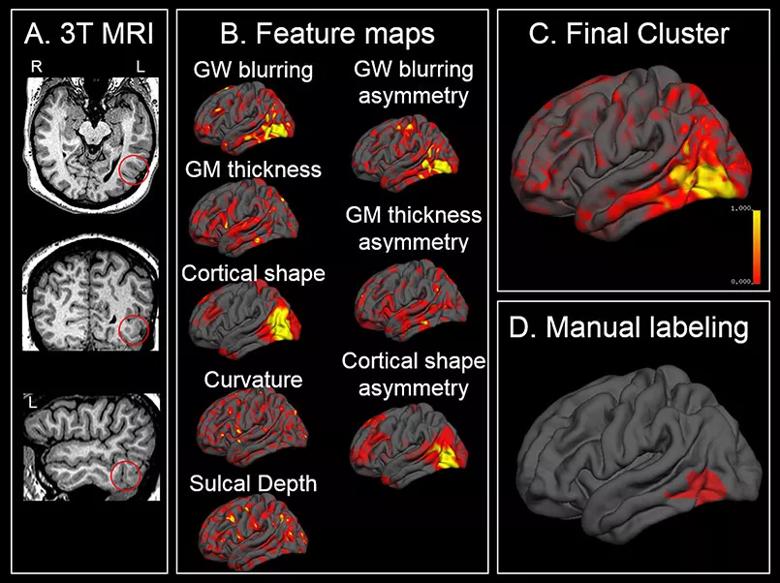
Image content: This image is available to view online.
View image online (https://assets.clevelandclinic.org/transform/4ba75ad9-082a-4e30-b3ea-4bb15266f99e/18-NEU-508-Nakamura-MRI-inset4_jpg)
Figure 4. Images illustrating a multivariate surface-based morphometry approach for automated focal cortical dysplasia (FCD) detection. (A) 3T T1w MRI from a patient with histopathologically confirmed type I FCD in the left basal temporal area (circles). (B) Various cortical features generated from the surface-based morphometry approach, which were used as input to a nonlinear neural network classifier. The output cluster with the highest mean probability value (as yielded by the classifier) was considered the final cluster (C). Success of detection was defined by overlap between the final cluster and manual labeling (informed by noninvasive test, SEEG, postoperative MRI and histopathology) (D).
Eventually, machine-learning techniques will enable the construction of a well-trained computer algorithm based on retrospectively validated data derived from a large number of patients. Such an algorithm could be used as an independent “consultant” to inform clinical recommendations on a case-by-case basis.
This strategy will have lasting impact across the country by markedly improving epilepsy seizure outcomes and increasing the number of patients who can be deemed favorable candidates for potentially curative surgery.
Advertisement
Dr. Wang (wangi2@ccf.org) is a staff scientist in Cleveland Clinic’s Epilepsy Center and joint staff in the Department of Biomedical Engineering, Cleveland Clinic Lerner Research Institute.
Dr. Nakamura (nakamuk@ccf.org) is a project scientist in the Department of Biomedical Engineering.
Advertisement
Advertisement

New study advances understanding of patient-defined goals

Testing options and therapies are expanding for this poorly understood sleep disorder

Real-world claims data and tissue culture studies set the stage for randomized clinical testing

Digital subtraction angiography remains central to assessment of ‘benign’ PMSAH

Cleveland Clinic neuromuscular specialist shares insights on AI in his field and beyond

Findings challenge dogma that microglia are exclusively destructive regardless of location in brain

Neurology is especially well positioned for opportunities to enhance clinical care and medical training

New review distills insights from studies over the past decade