Locations:

Experts to gather in Cleveland March 11-14
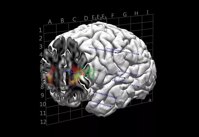
Image content: This image is available to view online.
View image online (https://assets.clevelandclinic.org/transform/e1f79d77-c893-4bc2-af8a-7af3187e3173/19-NEU-5567-Artificial-Intelligence-Epilepsy-Presurgical-Evaluation_jpg)
brain mapping in epilepsy
Effective use of stereoelectroencephalography (SEEG) for refractory epilepsy requires an accurate implantation strategy to ultimately enable a successful resective surgery. Providing detailed expert guidance in developing such a strategy is the overarching goal of Cleveland Clinic’s Ninth Brain Mapping Workshop, to take place in Cleveland from March 11 to 14, 2020.
Advertisement
Cleveland Clinic is a non-profit academic medical center. Advertising on our site helps support our mission. We do not endorse non-Cleveland Clinic products or services. Policy
The CME-certified course, titled “Imag(in)ing SEEG With Multimodal Integration,” will address the pre-SEEG workup — with multimodal guidance from MRI, EEG, MEG, SPECT and PET — as well as how multimodal findings are best used to inform surgical strategy development.
“A method for interpreting noninvasive multimodal data obtained during the planning phase of SEEG is essential to its success,” says Juan Bulacio, MD, an epileptologist with Cleveland Clinic’s Epilepsy Center who serves as one of the course’s five co-directors. “This procedure is not just for reviewing findings from different techniques. A fundamental interpretation is needed to integrate the various noninvasive findings according to their significance and their time sequence in the epileptogenic process. This brain mapping workshop will explore methods for doing so.”
The workshop is intimate in design, featuring opportunities for hands-on training and interaction with tutors dedicated to various case-based learning exercises. The faculty consists of epileptologists, neurosurgeons, neuroimaging specialists and neurophysiologists from Cleveland Clinic and other leading U.S. and European centers.
After kicking off Wednesday evening, March 11, with two hour-long introductory sessions on multimodality imaging and SEEG, the workshop structures most of the rest of its agenda around stages of the epileptogenic process:
Advertisement
After a Friday evening reception, the event concludes with a 3.5-hour Saturday morning session in which renowned epilepsy experts present in-depth cases for application of the workshop’s preceding insights.
Participants can expect to come away with a clear understanding of how multimodal studies contribute to the pre-SEEG workup as well as how to integrate the neurophysiological assessment for that workup to define a surgical strategy.
“Attendees will be able to interpret SEEG recordings and cortical stimulation findings in the context of the pre-SEEG multimodal imaging data,” says course co-director Irene Wang, PhD, a staff scientist in Cleveland Clinic’s Epilepsy Center. “Emphasis will be put on linking 2D data to 3D representation; this is crucially import for the interpretation of SEEG, which is 3D in nature.”
For registration and more information, visit ccfcme.org/brainmapping20.
Advertisement
Advertisement

New study advances understanding of patient-defined goals

Testing options and therapies are expanding for this poorly understood sleep disorder

Real-world claims data and tissue culture studies set the stage for randomized clinical testing

Digital subtraction angiography remains central to assessment of ‘benign’ PMSAH

Cleveland Clinic neuromuscular specialist shares insights on AI in his field and beyond

Findings challenge dogma that microglia are exclusively destructive regardless of location in brain

Neurology is especially well positioned for opportunities to enhance clinical care and medical training

New review distills insights from studies over the past decade