Locations:

Microscopy reveals surprising origin of attack on myelin membrane in MS model
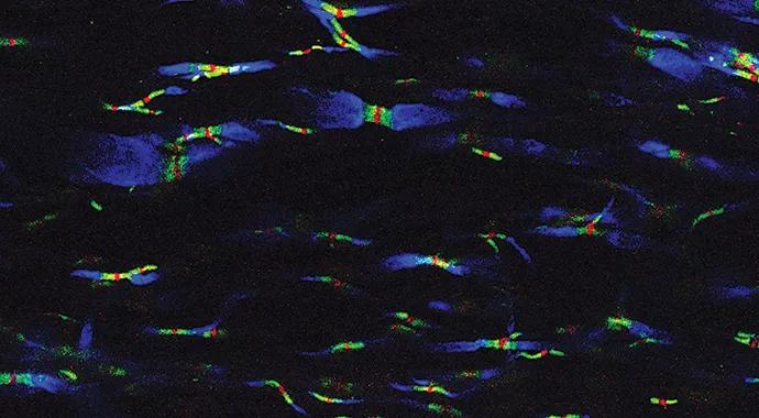
Image content: This image is available to view online.
View image online (https://assets.clevelandclinic.org/transform/1386db58-ffa8-402b-94cd-13f6802cdb75/ransohoff-690x380_jpg)
ransohoff-690×380
By Richard M. Ransohoff, MD, and Haiyan Lu, MD, PhD
Advertisement
Cleveland Clinic is a non-profit academic medical center. Advertising on our site helps support our mission. We do not endorse non-Cleveland Clinic products or services. Policy
Recent investigations by a Cleveland Clinic-led multicenter research team are yielding high-resolution representations of how inflammatory demyelination — the process underlying multiple sclerosis (MS) — begins. This article reviews the rationale behind this research, the essentials of our findings so far, and a glimpse ahead to potential implications and our next research goals.
In its simplest form, inflammatory demyelination results from the generation of autoimmune T lymphocytes, which can be stimulated by fragments of myelin, the fatty/proteinaceous membrane wrapped around nerve fibers. Once stimulated, the T cells communicate to macrophages, which then carry out the direct attack on myelin membranes. This attack results in patches of damage to myelin and nerve fibers, and this damage underlies the symptoms of MS.
The myelin membrane carries responsibility for ensuring energy-efficient, accurate communication among nerve cells as well as guaranteeing nourishment to nerve fibers. It is therefore critically important for proper nervous system function. Myelin damage can be repaired through endogenous recovery mechanisms, but this repair process is uncertain. As a result, prevention of myelin damage is a priority of MS research.
Despite nearly a century of research and much gratifying, clinically relevant progress, certain fundamental questions about inflammatory demyelination remain unresolved. A major engine driving MS research involves a similar disease, experimental autoimmune encephalomyelitis (EAE), which can be induced in mice by an immunization procedure that makes the mice “allergic” to their own myelin. About 2.5 weeks after the immunization, mice develop hind limb weakness very similar to the limb weakness experienced by MS patients. Several currently used MS treatments emerged directly from EAE studies.1
Advertisement
Using this EAE model, our Cleveland Clinic-led multicenter research team set out to evaluate how macrophages attack myelin during EAE in the mouse in a newly published study.2 We used serial block-face scanning electron microscopy (SBFSEM) with three-dimensional (3-D) reconstruction to make pictures of macrophages attacking myelin at micrometer (millionths of a meter) resolution.
These images showed a dramatic, unexpected representation of how inflammatory demyelination begins — namely, with macrophages being attracted to nodes of Ranvier. The nodes of Ranvier are basic to the structure of nerve fibers: They are the 1-μm gaps between myelin segments, where one myelin segment ends and the next begins (Figure 1). It was surprising and provocative to find macrophages localizing to these gaps in the myelin sheath. Our interest was strongly engaged because this feature of inflammatory demyelination might provide clues about the molecular signals that attract macrophages.
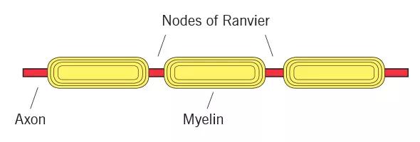
Image content: This image is available to view online.
View image online (https://assets.clevelandclinic.org/transform/9dec335d-bff3-4cc8-9940-a077dfa6696a/ransohoff-fig1_jpg)
Figure 1. Our recent study revealed that nodes of Ranvier, the 1-μm gaps between myelin segments along the axon, are where macrophages localize in experimental autoimmune encephalomyelitis, an animal model of MS.
We set about to develop a method for visualizing the nodes of Ranvier, both in healthy and in inflamed mouse spinal cord sections. We used confocal microscopy, which allowed us to focus on a flat focal plane less than 0.5 μm thick. This approach enabled us to see very small structures at quite high resolution. We performed pilot studies using dozens of tissue markers and many different ways of preparing spinal cord tissues for our studies.
Advertisement
Finally, we arrived at our goal: sharp, clear pictures showing nodes of Ranvier and the adjacent myelin structures in mouse spinal cord, from both healthy and inflamed tissues (Figure 2).
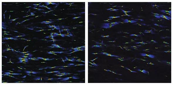
Image content: This image is available to view online.
View image online (https://assets.clevelandclinic.org/transform/f536e0e4-cd4b-4025-ad1d-1dd25c203dad/ransohoff-fig2_jpg)
Figure 2. Confocal microscopy images showing nodes of Ranvier in the spinal cords of a healthy mouse (left) and a mouse at the earliest stage of EAE (right). The blue, green and red structures in the left panel show the nodal elements as they should appear. The right panel shows swelling of the blue structures and lengthening of the green structures in some nodes. We hypothesize that these changes may expose markers recognized by invading macrophages.
We are now ready to embark on our next adventure: illuminating the macrophages as they attack these tissues and searching for their molecular targets. Stay tuned.
Dr. Ransohoff recently retired from long-standing appointments in Cleveland Clinic’s Neurological Institute and Lerner Research Institute to continue in the private sector his research into the pathogenesis and treatment of neurological disease. He remains Adjunct Professor of Molecular Medicine at Cleveland Clinic Lerner College of Medicine.
Dr. Lu is a research associate in Cleveland Clinic’s Department of Neurosciences.
Advertisement
Advertisement

New study advances understanding of patient-defined goals

Testing options and therapies are expanding for this poorly understood sleep disorder

Real-world claims data and tissue culture studies set the stage for randomized clinical testing

Digital subtraction angiography remains central to assessment of ‘benign’ PMSAH

Cleveland Clinic neuromuscular specialist shares insights on AI in his field and beyond

Findings challenge dogma that microglia are exclusively destructive regardless of location in brain

Neurology is especially well positioned for opportunities to enhance clinical care and medical training

New review distills insights from studies over the past decade