Locations:

Insights from the initial Cleveland Clinic experience
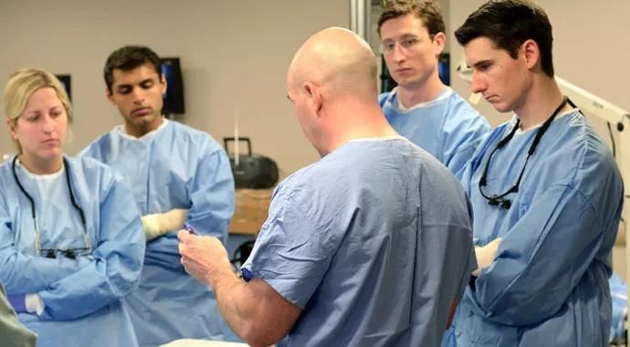
Image content: This image is available to view online.
View image online (https://assets.clevelandclinic.org/transform/47a29b29-4442-4bb9-bde9-27fd5a1b0747/kshettry-690x380_jpg)
kshettry-690×380
By Varun R. Kshettry, MD, and Richard Schlenk, MD
Advertisement
Cleveland Clinic is a non-profit academic medical center. Advertising on our site helps support our mission. We do not endorse non-Cleveland Clinic products or services. Policy
Resident training faces a multitude of changes. Duty hour restrictions impose limits on training time and the total number of cases performed by residents while in training. Given the lack of a corresponding decrease in clinical workload, there remains less time for educational activities. Additionally, healthcare system evolution has reduced resident autonomy in the operating room (OR) compared with what staff experienced during their training.
To adapt to these changes and maintain a high standard for our graduating trainees, educators are being forced to devise smarter educational techniques. We can no longer rely on the “sponge” educational concept ‒ i.e., if trainees spend enough time in the hospital and OR, they will absorb enough to reach a critical threshold in knowledge base and surgical technique.
Following a model from the aviation industry, other specialties, such as general surgery and urology, have adopted simulator training.1-5 However, computer simulation is better suited to on-screen modalities such as laparoscopy and endovascular therapy. Although some preliminary spine simulators have been developed for screw placement, these simulators leave something to be desired in terms of realism, anatomic detail and translation to the OR.6
We believe there is no substitute for hands-on experience. Visuospatial skills and muscle memory cannot be developed through reading. In the OR, residents do not have the freedom afforded by the laboratory environment. In the current healthcare climate, there is greater emphasis on reducing OR time, so trainee education is balanced against maintaining flow in the OR. Additionally, the laboratory affords opportunities to take surgical risks that cannot be taken in the OR due to the spine’s proximity to critical neurovascular structures.
Advertisement
In 2011, we began a formal educational dissection curriculum for Cleveland Clinic neurosurgery residents in postgraduate years (PGY) 2 through 5. Time was blocked on Friday afternoons once a month for six residents to leave their clinical duties to attend. We alternated between sessions for PGY 2-3 and PGY 4-5 residents in order to tailor activities to different baseline knowledge and skill levels.
Starting from scratch in early 2010, we needed about a year to secure laboratory space, equipment and funding for specimens. Funding was obtained largely from industry, with smaller contributions from the institution. Care was taken to structure agreements with industry for grant money to be given to the department for educational use only, free from any industry influence and in accordance with current guidelines for physician industry interactions. Because of initial space and budget limitations, the curriculum’s first year covered solely skull base surgery approaches. In 2012-2013, we incorporated a focused curriculum for spine and peripheral nerve surgery.
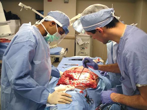
Image content: This image is available to view online.
View image online (https://assets.clevelandclinic.org/transform/08dae71a-1830-47b3-a6a3-201613dcb4a0/kshettry-inset_jpg)
While nadvantages of an Institutionally Based Lab Curriculumtional courses provide a reasonable alternative, there are several key advantages of an institutionally based laboratory curriculum:
Advertisement
We certainly recognize the hurdles in forming such a curriculum, including the need to secure instrumentation, lab space and specimens. Faculty interest and availability are also prerequisites, as is resident time away from clinical duties. For centers that find these barriers too burdensome, courses remain a good second option.
The formation of a standardized or suggested trainee dissection curriculum by governing organizations would help training programs during the start-up phase. A dissection guide would also serve as a good educational resource for residents during their training.
Given the overwhelmingly positive response from our residents, we plan to expand the number of sessions from 12 a year to 14 to 18 a year. Other plans include:
Incorporating a formal spine and peripheral nerve dissection curriculum into our neurosurgical training program has been a resounding success. Establishing a similar program involves overcoming multiple barriers, including the need to obtain space, equipment, funding for specimens, and resident and faculty availability. We believe a formal lab dissection curriculum can augment training in ways that simulators and courses cannot. Development of suggested curriculum or dissection guides focused on resident trainees would ease the transition.
Dr. Kshettry is a resident physician in the Department of Neurological Surgery and Resident Director of the Neurosurgical Skills and Anatomy Laboratory.
Advertisement
Dr. Schlenk is a spine surgeon in the Center for Spine Health and Director of the Neurosurgery Residency Program.
1. Gurusamy KS, Aggarwal R, Palanivelu L, Davidson BR. Virtual reality training for surgical trainees in laparoscopic surgery. Cochrane Database Syst Rev. 2009;(1):CD006575.
4. van Dongen KW, Ahlberg G, Bonavina L, et al. European consensus on a competency-based virtual reality training program for basic endoscopic surgical psychomotor skills. Surg Endosc. 2011;25(1):166-171.
5. Verdaasdonk EG, Dankelman J, Lange JF, Stassen LP. Transfer validity of laparoscopic knot-tying training on a VR simulator to a realistic environment: a randomized controlled trial. Surg Endosc. 2008;22(7):1636-1642.
Advertisement
Advertisement

New study advances understanding of patient-defined goals

Testing options and therapies are expanding for this poorly understood sleep disorder

Real-world claims data and tissue culture studies set the stage for randomized clinical testing

Digital subtraction angiography remains central to assessment of ‘benign’ PMSAH

Cleveland Clinic neuromuscular specialist shares insights on AI in his field and beyond

Findings challenge dogma that microglia are exclusively destructive regardless of location in brain

Neurology is especially well positioned for opportunities to enhance clinical care and medical training

New review distills insights from studies over the past decade