Locations:

How it adds value in structural assessment of bioprosthetic MV function
Advertisement
Cleveland Clinic is a non-profit academic medical center. Advertising on our site helps support our mission. We do not endorse non-Cleveland Clinic products or services. Policy
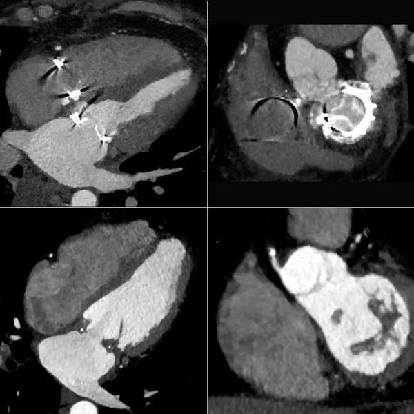
Image content: This image is available to view online.
View image online (https://assets.clevelandclinic.org/transform/9a1ab1a4-842b-453a-b573-c09adcd62985/17-HRT-3918-Cremer-inset_jpg)
Echocardiography is the mainstay for assessing bioprosthetic mitral valve function, but when it comes to assessing structure, there’s an emerging role for four-dimensional (4-D) CT. That’s particularly the case when there’s a diagnostic question of whether the predominant pathology is calcification versus valve thrombosis.
The 4-D CT images above highlight this point. The top two images are from a patient with symptomatic bioprosthetic mitral valve stenosis. On the left, the reconstructed apical four-chamber view shows hyperattenuated bioprosthetic valve leaflets, representing severe calcification. On the right, the short-axis reconstruction also shows leaflet hyperattenuation, consistent with calcification.
In contrast, the bottom two images are from a case of subacute bioprosthetic valve thrombosis in a patient with symptomatic mitral stenosis. On the left, the apical four-chamber reconstruction shows layering hypoattenuation along the sewing ring, indicating thrombus. On the right, the short-axis reconstruction shows prominent hypoattenuation and increased leaflet thickness illustrating the extension of thrombus onto the leaflets.
Imaging calcium is a strength of CT and a relative weakness of echocardiography, despite the overall strength of echo to assess the severity of bioprosthetic mitral valve dysfunction. The above images highlight how helpful 4-D CT can be in evaluating structural causes of bioprosthetic mitral valve dysfunction, particularly for distinguishing between calcification and thrombus.
Advertisement
Dr. Cremer (cremerp@ccf.org) is a cardiologist in Cleveland Clinic’s Section of Cardiovascular Imaging in the Sydell and Arnold Miller Family Heart, Vascular and Thoracic Institute.
For more reading on how 4-D imaging informs complex aortic valve repair in adult and pediatric patients, read this story from Cleveland Clinic Children’s of how a congenital heart surgeon and heart anatomist-imager team up to improve surgical success.
Advertisement
Advertisement

A sampling of outcome and volume data from our Heart & Vascular Institute

Concomitant AF ablation and LAA occlusion strongly endorsed during elective heart surgery

Large retrospective study supports its addition to BAV repair toolbox at expert centers

Young age, solid tumor, high uptake on PET and KRAS mutation signal risk, suggest need for lobectomy

Surprise findings argue for caution about testosterone use in men at risk for fracture

Residual AR related to severe preoperative AR increases risk of progression, need for reoperation

Findings support emphasis on markers of frailty related to, but not dependent on, age

Provides option for patients previously deemed anatomically unsuitable