Locations:

A new window into complex, highly variable anatomy
The tricuspid valve is a complex, highly variable structure that has been historically challenging to image with transesophageal echocardiography (TEE). As percutaneous options for tricuspid valve interventions have increased, so has the need for high-quality tricuspid imaging for use in preprocedural planning and intraprocedural guidance.
Advertisement
Cleveland Clinic is a non-profit academic medical center. Advertising on our site helps support our mission. We do not endorse non-Cleveland Clinic products or services. Policy
Cleveland Clinic is using state-of-the-art three-dimensional (3D) TEE to obtain high-resolution images to detail tricuspid anatomy. The number of leaflets, orientation and subvalvular apparatus can be identified clearly, as demonstrated in the representative images below.
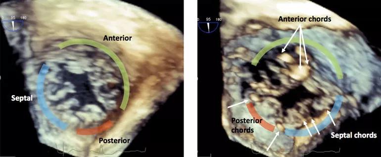
Image content: This image is available to view online.
View image online (https://assets.clevelandclinic.org/transform/f9a45b8a-a8aa-4e11-a2eb-83e7cb1c73e5/19-HRT-168-MiyasakaTEE-inset1-770x317_jpg)
3D TEE images showing atrial (left) and ventricular (right) views of the tricuspid valve in systole.
Furthermore, the use of live 3D multiplanar reconstruction has allowed us to provide highly detailed and accurate simultaneous 2D and 3D imaging for intraprocedural guidance of transcatheter tricuspid valve interventions, as reflected in the sample images below.
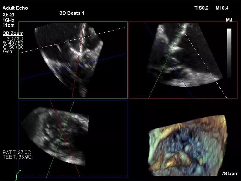
Image content: This image is available to view online.
View image online (https://assets.clevelandclinic.org/transform/f933ec6d-a8e2-4d29-ad68-b3c54020a2e4/19-HRT-168-MiyasakaTEE-inset2-770x579_jpg)
Live 3D multiplanar reconstruction allows for accurate real-time 3D image guidance.
For patients, 3D TEE technology translates into provision of the highest-quality imaging available for both the diagnosis and treatment of complex valvular heart disease.
Other recent innovations in tricuspid valve care include the use of 3D-printed models, isolated tricuspid valve surgery for right heart failure and the first implantation of the TricValve® Transcatheter Bicaval Valves System. For videos of note, watch the operative highlights from a tricuspid valve reconstruction for infective endocarditis or a summary of tricuspid valve percutaneous replacement and repair as a top medical innovation in 2019.
Images and text supplied by Rhonda Miyasaka, MD, a staff physician in Cleveland Clinic’s Section of Cardiovascular Imaging in the Sydell and Arnold Miller Family Heart, Vascular and Thoracic Institute.
Advertisement
Advertisement

A sampling of outcome and volume data from our Heart & Vascular Institute

Concomitant AF ablation and LAA occlusion strongly endorsed during elective heart surgery

Large retrospective study supports its addition to BAV repair toolbox at expert centers

Young age, solid tumor, high uptake on PET and KRAS mutation signal risk, suggest need for lobectomy

Surprise findings argue for caution about testosterone use in men at risk for fracture

Residual AR related to severe preoperative AR increases risk of progression, need for reoperation

Findings support emphasis on markers of frailty related to, but not dependent on, age

Provides option for patients previously deemed anatomically unsuitable