Locations:

Uncommon presentation with preseptal and nasal swelling
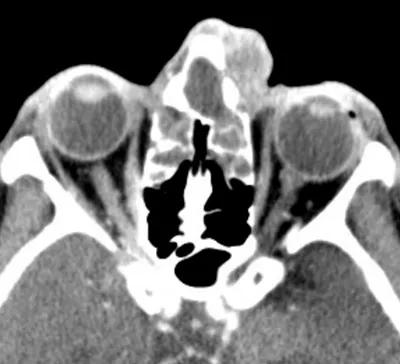
Image content: This image is available to view online.
View image online (https://assets.clevelandclinic.org/transform/4a757451-5b89-4a46-8264-cb1a29d1e2b1/16-EYE-2531-Potts-CQD-Inset-2_jpg)
16-eye-2531-potts-cqd-inset-2
A 77-year-old female with a history of chronic sinusitis presented with left periorbital pain for five days. The patient showed preseptal and left nasal swelling upon examination (Image A). She was given oral antibiotics to combat the cellulitis.
Advertisement
Cleveland Clinic is a non-profit academic medical center. Advertising on our site helps support our mission. We do not endorse non-Cleveland Clinic products or services. Policy
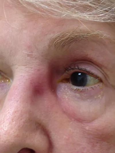
Image content: This image is available to view online.
View image online (https://assets.clevelandclinic.org/transform/b8b86887-45e9-45f5-93fd-f8534f8249fc/16-EYE-2531-Potts-CQD-Inset-1_jpg)
Imate A: Cellulitis in medial canthal region
Despite compliance with these antibiotics, the patient’s condition did not improve. Instead, she saw a dramatic increase in swelling five days after she started oral antibiotics (Image B).
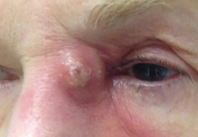
Image content: This image is available to view online.
View image online (https://assets.clevelandclinic.org/transform/9afb3984-7061-4592-9c76-2834765f31a0/16-EYE-2531-Potts-CQD-650x450_jpg)
Image B: Frontal sinus abscess
Julian Perry, MD, staff ophthalmologist at Cole Eye Institute, explained, “This patient presented with this uncommon entity in our outpatient service. Externally, it looked like acute dacryocystitis. However, the slightly anterior nature of the abscess increased our suspicion for a process arising from the frontal sinus.”

Image content: This image is available to view online.
View image online (https://assets.clevelandclinic.org/transform/4a757451-5b89-4a46-8264-cb1a29d1e2b1/16-EYE-2531-Potts-CQD-Inset-2_jpg)
Image C: CT showing dense opacification of frontal sinus with anterior table erosion.
A CT scan was ordered.
CT imaging revealed a destructive infection within the frontal sinus, causing some erosion of the anterior table. The abscess extended along the inferior portion of the frontal sinus (Image C). Pott’s puffy tumor typically manifests with swelling on the forehead associated with the frontal sinusitis and subperiosteal abscess. However, the patient’s symptoms and the CT imaging confirmed Dr. Perry’s theory that she had a rare presentation of Pott’s puffy tumor.
The patient’s symptoms were resolved with the combination of endoscopic treatment and extended intravenous antibiotics.
These images originally appeared in: Ganapathy P, Chundry R, Perry JD. Pott’s puffy tumor: a rare presentation. Ophthal Plast Reconstr Surg. 2016 Aug 16. [Epub ahead of print]. Reproduced with permission from Wolters Kluwer.
Advertisement
Advertisement
Early data shows risk is 73% higher in patients with lupus, 40% higher in patients with rheumatoid arthritis

Identifies weak spots in the cornea before shape change occurs

Study highlights the value of quantitative ultra-widefield angiography

Switching medications may decrease treatment burden and macular fluid

Interventions abound for active and stable phases of TED

Corneal imaging and interpretation play a major role
Cole Eye Institute imaging specialists are equal parts technician, artist and diagnostician

Effect of low-dose atropine and dual-focus contact lenses is unknown in patients with comorbid eye conditions