Locations:

Two cases showcase its versatile applications
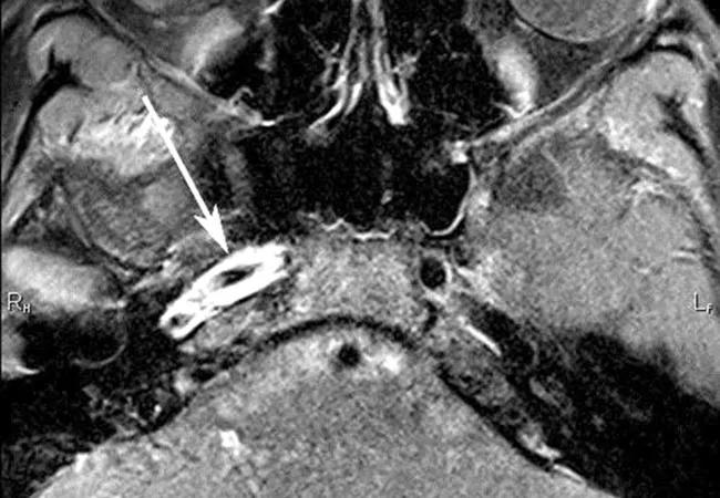
Image content: This image is available to view online.
View image online (https://assets.clevelandclinic.org/transform/5f10cd75-00d3-40c0-80fd-53ba02a03056/15-NEU-2533-Ruggieri-650x450_jpg)
15-NEU-2533-Ruggieri-650×450
Advertisement
Cleveland Clinic is a non-profit academic medical center. Advertising on our site helps support our mission. We do not endorse non-Cleveland Clinic products or services. Policy
Stroke is among the top 10 causes of pediatric mortality in the U.S. and results in significant morbidity in over half of survivors. High-resolution magnetic resonance (MR) vessel wall imaging helps identify abnormalities that result in either hemorrhagic or ischemic strokes. Identification of a cerebral vasculopathy in pediatric stroke is critical, in light of the increased risk of stroke recurrence in these cases.
Both MRI and MR angiography offer the advantage of noninvasive disease characterization without radiation. By using a reduced field of view, contrast enhancement and saturation pulses to eliminate artifacts inside blood vessels, it is possible to obtain high-resolution MRIs that delineate blood vessel walls rather than indirectly studying the internal contours of a blood vessel by imaging the flowing blood. High-resolution MRIs typically are obtained using a 3.0-Tesla whole-body scanner.
The technique described here refers specifically to imaging designed to assess the blood vessel walls. A gadolinium-based contrast agent is infused into the patient, allowing for vessel wall enhancement. To eliminate enhancement of flowing blood, a saturation pulse is employed to cancel the signal of blood perfusing the brain. By creating a signal-nulled “black blood” appearance, we can distinguish abnormal wall enhancement from enhanced blood.
Physicians collaborate to identify cases that might benefit from further analysis with vessel wall imaging and to plan tailored treatment for several stroke subtypes, including central nervous system vasculitis, dissection, and focal cerebral arteriopathy of childhood.
Advertisement
For instance, it may be unclear what is blocking blood flow in a vessel, such as in the internal carotid artery of the 10-year-old ischemic stroke patient whose imaging studies are shown in Figure 1.
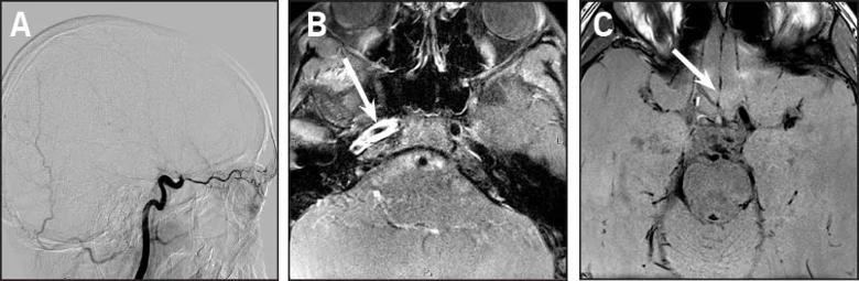
Image content: This image is available to view online.
View image online (https://assets.clevelandclinic.org/transform/751e30b7-2d13-4e2f-bce4-18b689efee6a/15-NEU-2533-Ruggieri-Fig1_jpg)
Figure 1. (First panel) Catheter angiogram showing blocked blood flow in the carotid artery of a 10-year-old patient with ischemic stroke. (Second panel) Axial contrast-enhanced T1 high-resolution MRI at the level of the petrous bone in the same patient showing proximal collapse of the vessel and luminal irregularity. (Third panel) Pre-contrast T1 high-resolution MRI of the patient’s blocked carotid artery showing high signal intensity along one side of the vessel wall, indicating blood beneath the intima of the wall and confirming a dissection as opposed to a thrombus.
The term vasculitides describes a diverse group of diseases that result in inflammation of a blood vessel wall, which may reduce blood flow to the brain area that the blood vessel supplies. High-resolution MRI can be used to identify the most diseased segments, potentially helping target blood vessels for biopsy. The technology can also help monitor response to treatment (see Figure 2).
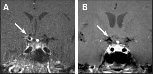
Image content: This image is available to view online.
View image online (https://assets.clevelandclinic.org/transform/a1f682f6-262b-4447-8400-7305d54ad193/15-NEU-2533-Ruggieri-Fig2_jpg)
Figure 2. These images show resolution of contrast enhancement along the walls of the right internal carotid artery and right middle cerebral artery origin on vessel wall imaging before and after treatment of varicella zoster virus-associated vasculitis.
Ryu C, Jahng G, Kim E, et al. High resolution wall and lumen MRI of the middle cerebral arteries at 3 Tesla. Cerebrovasc Dis. 2009;27(5):433-442.
Advertisement
Cheng-Ching E, Jones S, Hui FK, et al. High-resolution MrI vessel wall imaging in varicella zoster virus vasculopathy. J Neurol Sci. 2015;351(1-2):168-173.
Hui FK, Zhu X, Jones SE, et al. Early experience in high-resolution MRI for large vessel occlusions. J Neurointerv Surg. 2015;7(7):509-516.
Kuker W, Gaertner S, Nagele T, et al. Vessel wall contrast enhancement: a diagnostic sign of cerebral vasculitis. Cerebrovasc Dis. 2008;26(1):23-29.
Dr. Ruggieri is Section Head of MRI in Cleveland Clinic’s Imaging Institute and a staff member in Pediatric Neurosciences.
Advertisement
Advertisement

New study advances understanding of patient-defined goals

Testing options and therapies are expanding for this poorly understood sleep disorder

Real-world claims data and tissue culture studies set the stage for randomized clinical testing

Digital subtraction angiography remains central to assessment of ‘benign’ PMSAH

Cleveland Clinic neuromuscular specialist shares insights on AI in his field and beyond

Findings challenge dogma that microglia are exclusively destructive regardless of location in brain

Neurology is especially well positioned for opportunities to enhance clinical care and medical training

New review distills insights from studies over the past decade