Locations:

Imaging dye enables vascular assessment to promote procedural precision and safety
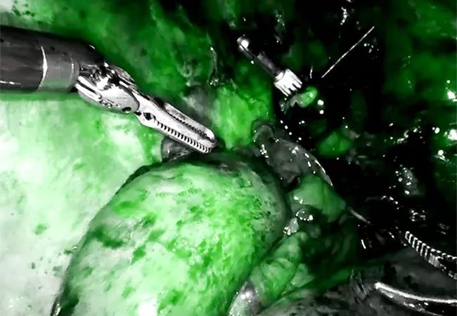
Image content: This image is available to view online.
View image online (https://assets.clevelandclinic.org/transform/790f9cac-6fe3-4e2f-b2fd-2847aa000989/23-HVI-3766947-CQD-650x450-1_jpg)
indocyanine green for vascular assessment during robotic esophagectomy
The thoracic surgery program in Cleveland Clinic’s Miller Family Heart, Vascular & Thoracic Institute performs some 250 to 300 robotically assisted operations each year. These include lobectomies, esophagectomies, operations for benign esophageal disease, and resection of mediastinal tumors.
Advertisement
Cleveland Clinic is a non-profit academic medical center. Advertising on our site helps support our mission. We do not endorse non-Cleveland Clinic products or services. Policy
Consistently high volumes make it easier for the program to adopt new technologies to improve operative safety and patient outcomes. One example is near-infrared intraoperative fluorescence imaging with the contrast agent indocyanine green (ICG), which Cleveland Clinic surgeons have integrated into a number of robotic thoracic surgery procedures.
The above image illustrates the use of ICG for vascular assessment of the gastric conduit during a robotically assisted esophagectomy in a patient with esophageal cancer. The presence of ICG (in green) serves to confirm that vascular perfusion of the conduit is adequate for reconstruction. “This gives us reassurance that we have good blood supply to the very tip of the stomach,” says Sudish Murthy, MD, PhD, Section Head of Thoracic Surgery at Cleveland Clinic. “It makes our operations safer for patients.”
Another thoracic surgery application of near-infrared intraoperative fluorescence imaging is in lung mapping for robotically assisted pulmonary resection. The contrast agent helps the surgeon identify which part of the lung is not perfused, allowing more precise targeting of the area that has been devascularized for resection.
Advertisement
Advertisement

A sampling of outcome and volume data from our Heart & Vascular Institute

Concomitant AF ablation and LAA occlusion strongly endorsed during elective heart surgery

Large retrospective study supports its addition to BAV repair toolbox at expert centers

Young age, solid tumor, high uptake on PET and KRAS mutation signal risk, suggest need for lobectomy

Surprise findings argue for caution about testosterone use in men at risk for fracture

Residual AR related to severe preoperative AR increases risk of progression, need for reoperation

Findings support emphasis on markers of frailty related to, but not dependent on, age

Provides option for patients previously deemed anatomically unsuitable