Locations:

As refinements continue, these advancements increasingly converge
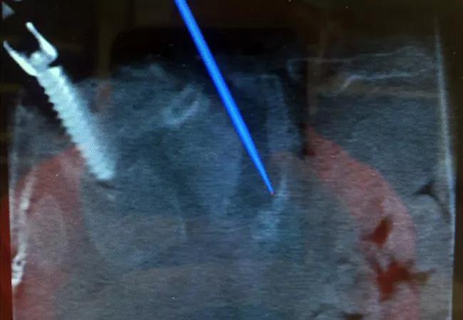
Image content: This image is available to view online.
View image online (https://assets.clevelandclinic.org/transform/883968f7-671d-460a-9ae8-f29b3cf84a58/17-NEU-4032-Savage-650x450_jpg)
17-NEU-4032-Savage-650×450
Advertisement
Cleveland Clinic is a non-profit academic medical center. Advertising on our site helps support our mission. We do not endorse non-Cleveland Clinic products or services. Policy
In the age of value-based healthcare, surgeons are constantly looking for ways to improve patient outcomes. Technological advancements and the desire to minimize the invasiveness of surgery have dramatically changed the way that spine surgery is performed in the 21st century.
The development of computer-assisted navigation systems has allowed surgeons to perform minimally invasive procedures, which are intended to improve outcomes and decrease direct and indirect healthcare costs. Image-guided navigation serves to improve the safety and ability to perform challenging procedures, including primary and metastatic tumor cases, revision surgery and complex spinal deformity reconstructions. Furthermore, spinal implants have dramatically improved over the past two decades. Surgeons now have a variety of biologics and biomaterials at their disposal, including 3-D-printed interbody cages, which serve to improve fusion rates in these often challenging healing environments.
This article briefly reviews these three advancements and how Cleveland Clinic’s Center for Spine Health and other leading centers are increasingly combining them in pursuit of better patient outcomes and care value.
Computer-assisted navigation has been around for many years, but the technology has improved over the past decade. Numerous intraoperative navigation systems are available to assist with surgery, particularly placement of pedicle screw instrumentation. While typically not used during routine spine cases, these navigation systems are often very helpful in revision surgeries and in complex deformities.
Advertisement
A recent systematic review (Gelalis et al, Eur Spine J. 2012;21:247-255) showed significantly improved accuracy of pedicle screw placement with the use of CT-based navigation systems. The percentage of screws fully contained in the pedicle ranged from 69 percent to 94 percent with a free-hand technique compared to 89 percent to 100 percent in the CT navigation cohort.
The use of navigation has also paved the way for performing less invasive surgical procedures, and robotic spine surgery is on the horizon.
Minimally invasive spine surgery (MISS) has been popularized and is now widely used for a variety of spinal disorders. MISS procedures aim to improve patient outcomes while decreasing the insult of surgery.
A wide array of MISS procedures are being performed in the U.S. and across the globe, including tubular discectomy/decompression, endoscopic discectomy, minimally invasive transforaminal interbody fusion, minimally invasive posterior cervical foraminotomy, lateral interbody fusion, percutaneous pedicle screw instrumentation and minimally invasive deformity correction. In general, MISS has been shown to be associated with less intraoperative blood loss, improved perioperative pain scores and time to ambulation, and decreased length of hospital stay (Goldstein et al, J Neurosurg Spine. 2016;24:416-427).
MISS procedures have also been shown to be cost-effective; however, MISS procedures have not yet been shown to improve long-term patient outcomes compared with traditional open techniques. Patient-reported outcomes are likely driven by surgical indications, proper technique and optimization of perioperative medical and psychosocial comorbidities — and may be less related to the size of the incision.
Advertisement
Lumbar fusion procedures are commonly performed in the setting of spinal instability (isthmic or degenerative spondylolisthesis and/or spinal deformity). Fusion rates have varied over the years, with a wide range of success, and it has been shown that achieving a solid fusion leads to improved long-term outcomes.
The recent introduction of new and improved biomaterials may improve fusion rates. New interbody fusion “cages” are being developed to improve the devices’ ability to integrate with bone. 3-D-printed cages are available that optimize the structural properties and porosity of the metal, which will in theory improve the “biologic” capability of the cages. The ultimate goal is to improve the osseous integration of the cage, which should improve fusion rates, in hope of resulting improvements in long-term patient outcomes.
At Cleveland Clinic, MISS procedures are performed routinely, and intraoperative navigation is often used to assist in complex revision and deformity operations, as demonstrated by the two cases presented in images below. Case 1 illustrates a minimally invasive transforaminal interbody fusion with the use of intraoperative navigation to place cortical screws and a 3-D-printed interbody cage. Case 2 illustrates the use of intraoperative CT navigation to place iliac fixation in a patient with a Charcot lumbosacral arthropathy.
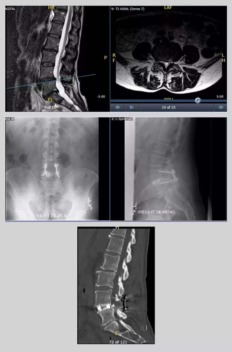
Image content: This image is available to view online.
View image online (https://assets.clevelandclinic.org/transform/480d7a9e-5514-480b-b223-cfe6f5060874/17-NEU-4032-Inset1_jpg)
Case 1. Images from the case of a 67-year-old woman with bilateral leg pain when standing and walking. Preoperative anteroposterior and lateral X-rays (top panel) show a grade 1 L4-L5 degenerative spondylolisthesis with lateral recess stenosis. Postoperative X-rays (middle panel) and CT scan (bottom panel) reveal good positioning of the interbody cage and cortical screws with solid arthrodesis.
Advertisement
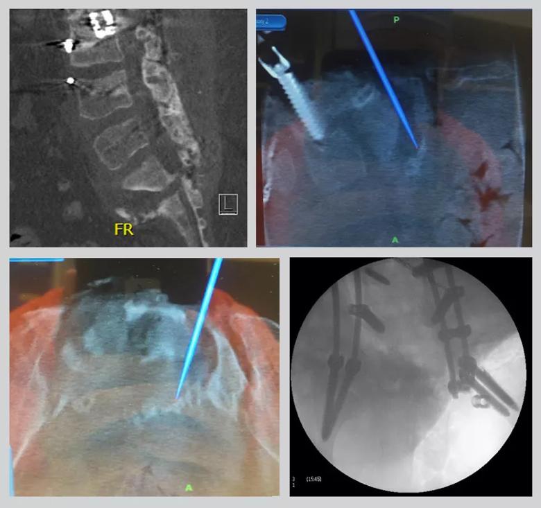
Image content: This image is available to view online.
View image online (https://assets.clevelandclinic.org/transform/16dd7fa4-cef0-49a4-a04d-e165c7fcc681/17-NEU-4032-Inset2_jpg)
Case 2. Images from the case of a 21-year-old woman with a history of paraplegia who developed a lumbosacral Charcot arthropathy after a T4-ileum reconstruction. Preoperative imaging studies (top left) show the Charcot arthropathy. Intraoperative navigation was used (top right and bottom left) to identify the extent of the bone loss and aid in the placement of lumbopelvic fixation. Intraoperative imaging (bottom right) shows the placement of four iliac screws.
The field of spine surgery continuously strives to improve patient outcomes. Technological advances have paved the way for computer-assisted and minimally invasive operations. Continued developments in osteobiologics and enhancements in biomaterials will hopefully improve fusion rates and, in turn, long-term patient outcomes. A concerted effort to prospectively evaluate these procedures, their outcomes and the cost-effectiveness of various surgical approaches is absolutely necessary as we proceed toward value-based spine care.
Dr. Savage is a surgeon and Spine Surgery Fellowship Director in Cleveland Clinic’s Center for Spine Health.
Advertisement
Advertisement

New study advances understanding of patient-defined goals

Testing options and therapies are expanding for this poorly understood sleep disorder

Real-world claims data and tissue culture studies set the stage for randomized clinical testing

Digital subtraction angiography remains central to assessment of ‘benign’ PMSAH

Cleveland Clinic neuromuscular specialist shares insights on AI in his field and beyond

Findings challenge dogma that microglia are exclusively destructive regardless of location in brain

Neurology is especially well positioned for opportunities to enhance clinical care and medical training

New review distills insights from studies over the past decade