Locations:

7T MRI scanners enable system-level brain study at near-microscopic scale
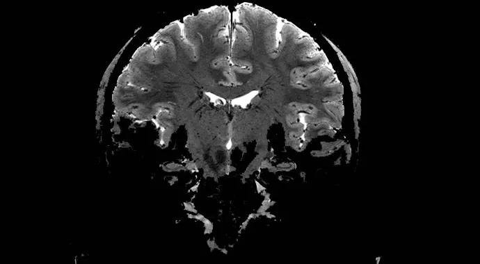
Image content: This image is available to view online.
View image online (https://assets.clevelandclinic.org/transform/37a5cef4-49e1-40b9-b58b-cf26cc9f1805/NI_hippo_hires_cor1-690_jpg)
NI_hippo_hires_cor1-690
Cleveland Clinic recently installed a 7-tesla MRI scanner on its main campus, making it one of only 13 medical centers in the United States with 7T MRI scanning abilities.
Advertisement
Cleveland Clinic is a non-profit academic medical center. Advertising on our site helps support our mission. We do not endorse non-Cleveland Clinic products or services. Policy
The new scanner’s more than doubling in field strength (relative to the previous 3T standard) permits researchers to produce in vivo body images at spatial resolutions up to five times greater than those possible at clinical field strengths. Its images can approach the spatial resolution of CT scans while maintaining the superior soft-tissue contrast of MRI.
Because of FDA restrictions on the clinical use of MRI scanners with field strengths above 4T, the new scanner will be used for biomedical research purposes.
“With this 7T imaging facility, we can begin to explore brain function and structure at an unprecedented level,” says Mark Lowe, PhD, Cleveland Clinic’s Director of High-Field MRI, whose team finalized installation of the scanner in December 2013.
“The advantage of MRI has always been the ability to study the brain as a system,” he explains. “This greatly enhances that ability by allowing us to simultaneously study the brain both as a system and at nearly microscopic scales.”
The new scanner’s enhanced imaging research capabilities are still being explored and prioritized. For one, functional MRI is likely to be improved, as the higher field strength will allow for its performance at very high spatial and temporal resolution, which will open up new areas of investigation in neuropsychiatry and other research areas.
“We will be working to develop magnetic resonance spectroscopic imaging (MRSI) sequences suited for ultra-high fields to take advantage of the increased spatial and spectral resolution they provide,” notes Amit Anand, MD, Vice Chairman for Research in Cleveland Clinic’s Center for Behavioral Health.
Advertisement
Extremely high-resolution diffusion tensor imaging of the hippocampus with the 7T scanner may permit discovery of novel biomarkers in Alzheimer disease and other cognitive disorders.
Areas of epileptogenic focus are normally too small to identify with standard MRI, but one hope is that these foci will be able to be localized with 7T imaging, enabling more precise surgical removal of seizure-producing brain areas.
When it comes to brain tumors, the high spatial resolution and contrast-to-noise ratio of 7T imaging will allow for superior definition of tumor boundaries within the brain. In musculoskeletal imaging, it has already been demonstrated that the higher field strength permits better imaging of cartilage.
Previously, all whole-body MRI scanners at Cleveland Clinic produced images based on proton excitation. Protons are the most abundant nuclei in the human body and by far the most common nuclei used to produce MRI images. However, other nuclei in the body occur naturally in a form that can be studied with MRI.
Beyond its high field strength, the new 7T system is the first whole-body MRI scanner at Cleveland Clinic with multinuclear imaging capability. In addition to producing images that provide anatomic or functional detail from protons, the scanner is able to yield information on metabolic processes using other nuclei, such as sodium, phosphorus and carbon. This capability permits the study of more complex processes, such as cell metabolism.
The 7T MRI scanner’s journey to Cleveland and installation were nearly as impressive as its imaging capabilities.
Advertisement
Installation, which included the opening of a 7T MRI facility adjacent to Cleveland Clinic’s Mellen Center for Multiple Sclerosis Treatment and Research, began in July 2013, and the scanner started producing images by September.
The magnet at the heart of the 7T system is constructed to order by its manufacturer, Siemens Healthcare, over 18 months. Behind this long production window is the enormous amount of superconducting wire — stretching 350 miles in length — needed to produce the magnet.
To maintain field strength, the wire must be kept very cold, requiring 20,000 liters of liquid helium for cooling. At 80,000 pounds, the magnet had to be shipped by boat from its production site in Oxford, England, taking three weeks to cross the Atlantic Ocean.
Because the helium would boil away during a journey of that length, the magnet was shipped warm and then cooled at Cleveland Clinic over several weeks. Another several weeks were needed to pump current through the magnet to get it up to field strength. The overall process was slowed by the worldwide helium shortage of the past few years.
Once the magnet was up to field strength, Siemens engineers needed 90 days to finish installation of the electronics necessary to produce high-quality images. Physicists from Cleveland Clinic’s Imaging Institute then performed final tuning to customize the scanner facility for the specific research needs desired.
Advertisement
Advertisement

New study advances understanding of patient-defined goals

Testing options and therapies are expanding for this poorly understood sleep disorder

Real-world claims data and tissue culture studies set the stage for randomized clinical testing

Digital subtraction angiography remains central to assessment of ‘benign’ PMSAH

Cleveland Clinic neuromuscular specialist shares insights on AI in his field and beyond

Findings challenge dogma that microglia are exclusively destructive regardless of location in brain

Neurology is especially well positioned for opportunities to enhance clinical care and medical training

New review distills insights from studies over the past decade