Locations:

May hold implications for full spectrum of neurologic disease
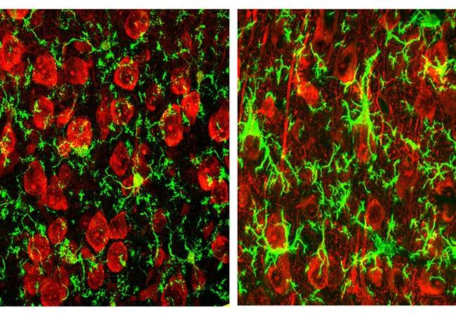
Image content: This image is available to view online.
View image online (https://assets.clevelandclinic.org/transform/f34d1554-c6bc-412d-8ede-19c183c6a8a2/15-NEU-219-Chen-650x450_jpg)
15-NEU-219-Chen-650×450
By Zhihong Chen, PhD, and Bruce D. Trapp, PhD
Advertisement
Cleveland Clinic is a non-profit academic medical center. Advertising on our site helps support our mission. We do not endorse non-Cleveland Clinic products or services. Policy
Resident macrophages exist in almost all organs, where they are the first-line defenders against infection or disease. In the brain, this innate immune response is performed by microglia. Numerous microglia cover the entire brain parenchyma in a nonoverlapping, mosaic fashion by spreading their delicate processes, as illustrated in the image above.
Modern multiphoton imaging techniques have allowed glial biologists to directly study the brain of the living mouse. What is astonishing is that microglial processes can be observed constantly extending or retracting a minute distance, as reported by Nimmerjahn and colleagues, making them arguably the fastest-moving structures (~1.5 μm/min) in the brain.
These observations aroused strong curiosity about what microglia actually do and why they do it — questions that remain to be fully addressed today. However, at least one hypothesis is that these movements allow microglia to actively gauge the health of surrounding central nervous system (CNS) cells within their microdomains.
Microglia are activated by acute insults and chronic diseases. This activation induces hypertrophy of their cell bodies, asymmetrical distribution of their processes and increased expression of activation molecules.
Activated microglia can be observed in many neurologic diseases, such as those surrounding the core plaques in an Alzheimer disease brain. Because of their frequent presence in various disease states, activated microglia traditionally have been considered to be destructive.
Advertisement
This “guilt by association” view has recently been revisited, however, as more and more studies have begun to demonstrate that microglia are actually essential defenders against many CNS diseases. This is particularly the case in chronic brain diseases, where the majority of microglia are activated. If these activated microglia were exclusively destructive, these conditions would not be “chronic” because microglia would have essentially destroyed much of the tissues.
As we noted in a recent review, even more convincing evidence supporting a neuroprotective role for microglia lies in conditions where microglial activation occurs in the absence of frank pathology, such as when “rod cells” encapsulate healthy-appearing neurons, as observed in subacute sclerosing panencephalitis, multiple sclerosis (MS) or amyotrophic lateral sclerosis.
In our previous work, we found that microglial activation, as a critical part of immune responses in the CNS, can be mediated by a preconditioning paradigm induced by injecting the gram-negative bacteria outer membrane component lipopolysaccharide (LPS). We further showed that LPS-induced microglial activation contributes to neuroprotection against experimental traumatic brain injury, as the brain lesion size is much smaller when microglia are activated by LPS. This novel finding has facilitated revision of prior perceptions of the role and mechanism of microglial activation in the brain.
In the wake of our above findings in 2012, we were interested in the specific actions that activated microglia exert on neurons in order to provide protection. Our continued investigations involved applying several novel technologies, including three-dimensional electron microscopy and wireless telemetric electrophysiology.
Advertisement
In a breakthrough observation published in Nature Communications in 2014, we demonstrated for the first time that microglia, when activated (Figure 1), migrate to and dislodge inhibitory synapses between neurons. This “synaptic stripping” increases neuronal firing activity and leads to a cascade of events that enhance survival of brain cells (Figure 2). We further demonstrated that these events are one of the underlying mechanisms by which activated microglia contribute to neuroprotection after traumatic injury.
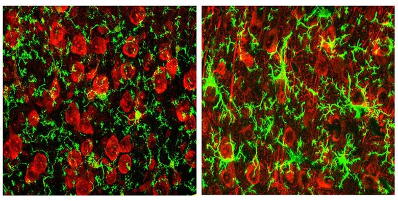
Image content: This image is available to view online.
View image online (https://assets.clevelandclinic.org/transform/bd628893-3c77-493c-b271-859a09d6fb0f/15-NEU-219-Chen-Inset-1_jpg)
Figure 1. Morphological appearance of microglia in the mouse brain (visualized in green by immunofluorescent staining with anti-Iba1 antibody). In control mice (left), microglia have small cell bodies and long and slender processes. When activated (right), microglia enlarge their cell bodies and thicken their processes, which closely enwrap neuronal cell bodies (red, Nissl staining). Reprinted from Chen et al, Nature Communications, ©2014, Nature Publishing Group.
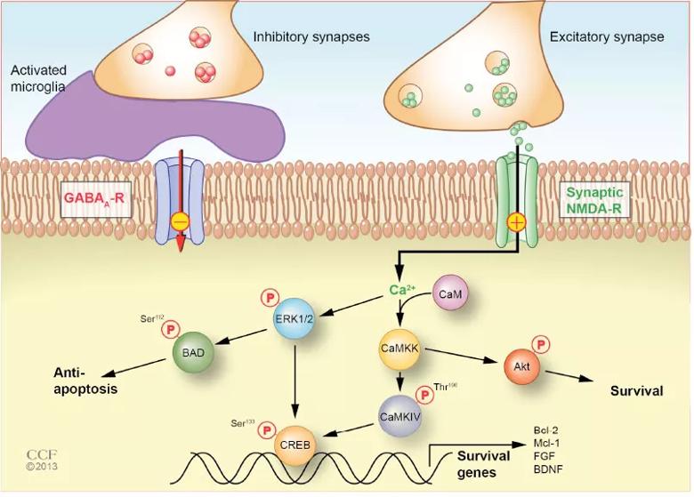
Image content: This image is available to view online.
View image online (https://assets.clevelandclinic.org/transform/d40ef072-87f6-4111-b7de-71b8b7a47fd2/15-NEU-219-Chen-Inset-2_jpg)
Figure 2. Model of activated microglia-mediated neuroprotection. Activated microglia displace presynaptic GABAergic terminals, which lowers the threshold for firing of excitatory synaptic NMDA receptors. Increased firing of synaptic NMDA receptors (+) elevates intracellular Ca2+ levels, which leads to activation (phosphorylation) of signaling molecules and transcription factors, culminating in production of anti-apoptotic and neurotrophic proteins. Reprinted from Chen et al, Nature Communications, ©2014, Nature Publishing Group.
Advertisement
Given that physical interactions between microglia and neurons exist in a variety of neurologic diseases, such as Alzheimer disease, MS and stroke, our discoveries may have a profound impact across the whole spectrum of neurologic disease. They suggest that the protective role of microglia could potentially be harnessed to improve the prognosis for patients with traumatic brain injury and delay the progression of diseases such as Alzheimer disease, MS and stroke.
On a broader scale, our findings suggest that the innate immune system helps protect the brain after injury or during chronic disease, and this role should be further studied.
Our lab is now working to elucidate the pathways and molecular mechanisms of microglial activation in the model we have established. We are using RNA microarray profiling techniques to define the molecular signature of neuroprotective microglia. Identifying the profile of the protecting microglia will aid the design of targeted therapeutic strategies.
Dr. Chen is a research associate in the Department of Neurosciences in Cleveland Clinic’s Lerner Research Institute.
Dr. Trapp is Chairman of the Department of Neurosciences in the Lerner Research Institute.
Advertisement
Advertisement

New study advances understanding of patient-defined goals

Testing options and therapies are expanding for this poorly understood sleep disorder

Real-world claims data and tissue culture studies set the stage for randomized clinical testing

Digital subtraction angiography remains central to assessment of ‘benign’ PMSAH

Cleveland Clinic neuromuscular specialist shares insights on AI in his field and beyond

Findings challenge dogma that microglia are exclusively destructive regardless of location in brain

Neurology is especially well positioned for opportunities to enhance clinical care and medical training

New review distills insights from studies over the past decade