Locations:

MOTION study will quantify new technology’s impact on radiation exposure
In December, Cleveland Clinic vascular surgeons successfully completed the first surgical case in the MOTION clinical trial of a new non-radiation-based surgical navigation platform for endovascular aortic interventions.
Advertisement
Cleveland Clinic is a non-profit academic medical center. Advertising on our site helps support our mission. We do not endorse non-Cleveland Clinic products or services. Policy
The trial is a feasibility study of the Intra-Operative Positioning System (IOPS™) from Centerline Biomedical, which received 510(k) marketing clearance from the FDA in 2019. The technology, which serves as an adjunct to X-ray fluoroscopy, is in use at Cleveland Clinic and four other U.S. institutions as part of a limited launch.
“The MOTION trial is being conducted to determine the degree of radiation reduction that’s possible with IOPS and to better characterize its utility in endovascular aortic repair [EVAR],” says Cleveland Clinic vascular surgeon Francis Caputo, MD, who performed the study’s first case and serves as its national principal investigator.
As detailed in an earlier Consult QD post, the IOPS technology was initially developed at Cleveland Clinic as a platform for enhancing visualization during endovascular aortic interventions with reduced reliance on fluoroscopy.
The portable system employs anatomic mapping algorithms and electromagnetic tracking technology to provide high-definition, three-dimensional (3D) color visualization and guidance in real time during endovascular procedures. “The technology is similar to GPS, in that it uses coordinates derived from prior imaging to guide navigation,” explains Dr. Caputo.
He says an approach like IOPS ultimately has potential to supplant fluoroscopy as the standard of care for visualization during EVAR procedures. “Not only does this system help guide accurate placement of catheters and guidewires as the surgeon navigates the aorta and its branches, but it has the potential to reduce harmful radiation exposure to both surgeons and patients through reduced dependence on fluoroscopy,” Dr. Caputo observes.
Advertisement
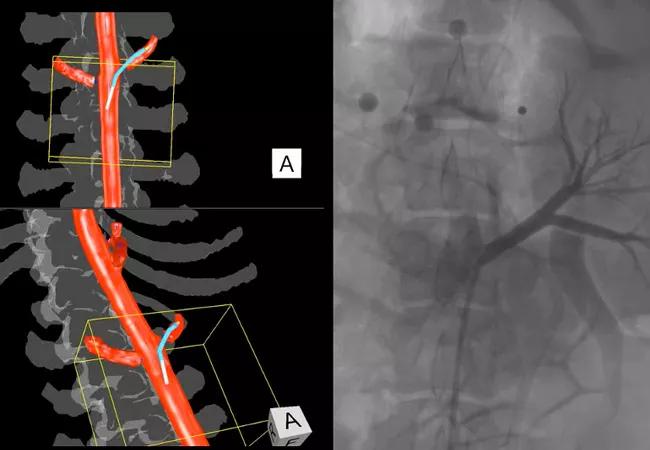
Image content: This image is available to view online.
View image online (https://assets.clevelandclinic.org/transform/4097aa31-39e0-4fb9-a5f0-12b4cb87ac94/20-HVI-2034258-EVAR-navigation_650x450_jpg)
Comparative visualization of the left renal artery with the IOPS platform (left) versus fluoroscopy and contrast dye (right).
In fact, radiation dose is one of the main measures being assessed in the MOTION trial, along with use of contrast dye and duration of the EVAR procedure. “IOPS was designed to reduce radiation exposure and use of toxic contrast dye, as well as reduce procedure time through enhanced visualization and coordinate-based navigation assistance,” says Dr. Caputo. “This study aims to formally quantify to what extent it achieves those objectives.”
MOTION is designed to enroll 30 patients between Cleveland Clinic and a second study site, UNC Medical Center in Chapel Hill, North Carolina. Completion of the single-arm study is anticipated by the end of 2021, after which a controlled study may be undertaken to compare IOPS plus fluoroscopy (as needed) versus fluoroscopy alone.
If MOTION confirms that IOPS enables substantial reductions in radiation dose, contrast use and procedure time, Dr. Caputo believes the technology is likely to be widely adopted for guiding EVAR procedures in the years ahead — particularly for longer, more complex cases that would otherwise require significant radiation exposure.
“The greatest potential from this system is in more complex cases, ones that involve considerable navigation to target vessels,” he explains. “With fluoroscopy alone, cases like that can require 5 to 10 gray of radiation, and levels above 5 gray raise concern about radiation risk to patients — as well as to providers, despite the lead shields we wear. Beyond safety benefits to patients and staff, reduced-radiation procedures may ultimately become an occupational safety requirement.”
Advertisement
After completing six EVAR repairs using IOPS through the end of 2020, Dr. Caputo says the system also offers advantages to the operator in terms of navigation. “For example,” he notes, “if you are targeting a renal artery, it shows you not only where the artery is but how to use the catheter and wire in conjunction to quickly home in on the artery. Achieving that type of navigation without much or any radiation is a long-sought objective that may be at hand.”
NOTE: Centerline Biomedical is a Cleveland Clinic spinoff company. Cleveland Clinic holds equity in the company and is entitled to royalties from the company.
Advertisement
Advertisement

A sampling of outcome and volume data from our Heart & Vascular Institute

Concomitant AF ablation and LAA occlusion strongly endorsed during elective heart surgery

Large retrospective study supports its addition to BAV repair toolbox at expert centers

Young age, solid tumor, high uptake on PET and KRAS mutation signal risk, suggest need for lobectomy

Surprise findings argue for caution about testosterone use in men at risk for fracture

Residual AR related to severe preoperative AR increases risk of progression, need for reoperation

Findings support emphasis on markers of frailty related to, but not dependent on, age

Provides option for patients previously deemed anatomically unsuitable