Locations:

Techniques help provide a complete picture of the coronary tree
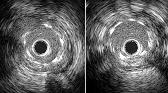
Image content: This image is available to view online.
View image online (https://assets.clevelandclinic.org/transform/5f95d842-f4bb-42ab-9c6e-fc764a9974e0/IVUS-690x380_jpg)
IVUS-690×380
Advertisement
Cleveland Clinic is a non-profit academic medical center. Advertising on our site helps support our mission. We do not endorse non-Cleveland Clinic products or services. Policy
While angiography remains the gold standard for diagnosing the presence and severity of a coronary artery lesion, it often does not tell the whole story. In some patients, an additional examination technique is needed to fully understand an area of the coronary tree that appears abnormal and help the clinician determine the need for treatment.
For these purposes, fractional flow reserve (FFR) and intravascular ultrasound (IVUS) are indispensible.
As a two-dimensional imaging modality, angiography has limitations, one of which is determining the hemodynamic significance of certain lesions. The value of FFR is backed by strong data. In these cases, FFR can be used to provide a functional assessment of the heart. In studies, FFR has been shown to be as effective as a thallium stress test in visualizing blood supply when demand is increased. Moreover, randomized, controlled trials have demonstrated that patients with FFR values >.80 can be managed safely without revascularization.
FFR is performed by passing a 0.014” wire carrying a pressure sensor through the lesion to obtain the distal pressure. This compliments information on the proximal pressure obtained during the catheterization and provides a baseline reading. We then give a vasodilator such as adenosine. Under normal conditions, no change in pressures is expected. Many times the pressure beyond the obstruction is lower. If the pressure beyond the narrowing is less than 80 percent that of the proximal reading, the FFR is abnormal. This indicates blood flow is obstructed, and the patient would benefit from revascularization.
Advertisement
At Cleveland Clinic, FFR is used only when it makes sense. When a patient has EKG changes and a 90 percent blockage, or a stress test reveals an inadequate blood supply, there is no need for FFR before proceeding with revascularization. But in patients with questionable symptoms and borderline tests, and in those with no stress test information, FFR can be very helpful.
In contrast to FFR, IVUS facilitates assessment of blood vessel morphology. The cross-sectional views provided by IVUS clearly delineate the structure of an artery wall and diameter of the lumen. We often use IVUS to better visualize arterial segments not well seen on angiography, such as near overlapping branches or branching points.
IVUS is also valuable for evaluation before and after stent placement, as well as research into the mechanism of in-stent restenosis.
A third technique called optical coherence tomography (OCT) is growing in importance. OCT provides extremely high-resolution images that reveal the nature and composition of a plaque. Experimental OCT devices can determine the molecular structure with astounding clarity.
OCT is proving to be a useful research tool for revealing the nature of atherosclerosis. The better we understand a given plaque, the better we know how threatening it is. Having this information enables us to choose the appropriate technique to prevent myocardial infarction.
Advertisement
Advertisement

A sampling of outcome and volume data from our Heart & Vascular Institute

Concomitant AF ablation and LAA occlusion strongly endorsed during elective heart surgery

Large retrospective study supports its addition to BAV repair toolbox at expert centers

Young age, solid tumor, high uptake on PET and KRAS mutation signal risk, suggest need for lobectomy

Surprise findings argue for caution about testosterone use in men at risk for fracture

Residual AR related to severe preoperative AR increases risk of progression, need for reoperation

Findings support emphasis on markers of frailty related to, but not dependent on, age

Provides option for patients previously deemed anatomically unsuitable