Locations:

Early effective intervention is key to improved QOL
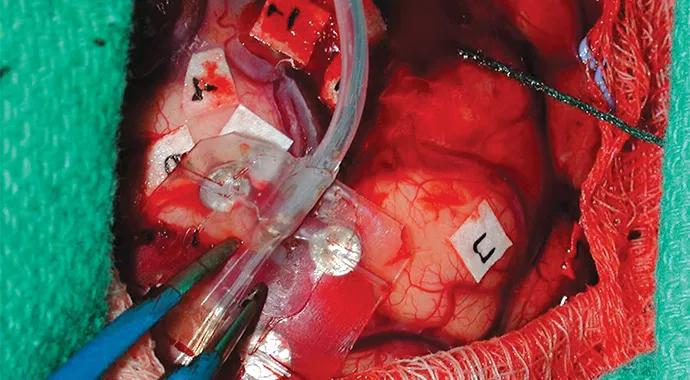
Image content: This image is available to view online.
View image online (https://assets.clevelandclinic.org/transform/66be87d5-a50b-4a45-8ef2-bce4203891e2/tuberous-selerosis-690x380_jpg)
tuberous-selerosis-690×380
By Ajay Gupta, MD
Advertisement
Cleveland Clinic is a non-profit academic medical center. Advertising on our site helps support our mission. We do not endorse non-Cleveland Clinic products or services. Policy
Tuberous sclerosis complex (TSC) is a genetic disease whose lifelong neurological morbidity is usually defined early in life by epilepsy and cognitive delay that typically go hand in hand. Early effective treatment of epilepsy is a key factor in improving quality of life in patients with TSC. Epilepsy surgery, despite its unique challenges in this population, offers hope for seizure freedom and enhanced quality of life in patients who fail to respond to medical treatment.
A successful surgical strategy in any patient with epilepsy involves identifying the epileptogenic zone that has the potential to be safely resected to obtain seizure freedom without risk of a new postoperative deficit. Challenges of epilepsy surgery in pediatric patients with TSC are more complex, but they are not insurmountable in the hands of experienced epilepsy specialists and surgeons working as a team.
The first challenge unique to TSC patients is to identify the culprit lesion (tubers and subcortical dysplasias) in the face of multiple lesions all over the brain. The investigation to find the culprit tuber — and define the epileptogenic zone — begins with a careful history and physical exam and a review of seizures on videos with simultaneous scalp EEG evaluation (video EEG) by an experienced epileptologist. In some patients, reviewing previous EEGs may offer crucial clues.
Brain MRI is a critical imaging tool for corroborating findings from the clinical and video EEG evaluation. Brain PET and ictal SPECT, although challenging in children with TSC, sometimes provide useful complementary data in selected children.
Advertisement
More recently, studies have found that magnetoencephalography and magnetic source imaging can be useful in identifying the epileptogenic tuber in patients with TSC.
The second challenge is to determine the safety of resecting the epileptogenic tuber if it is located close to the eloquent region for speech/language, vision, or motor or sensory control. When practical, newer techniques, such as functional MRI and MR tractography, may help clarify the surgical strategy in this situation.
In some cases invasive recordings may be necessary, such as a brain-mapping technique in which subdural grids and depth electrodes are placed to map the seizure onset zone and define its relationship to the area of the eloquent cortex. Such procedures often create a next level of complexity in infants and young children, due to lack of cooperation and electrophysiological immaturity of the central nervous system.
In some instances, invasive recordings can be accomplished in the operating room (intraoperative cortical stimulation and electrocorticography) under the safety of light anesthesia without the requirement of active patient cooperation (Figure 1).
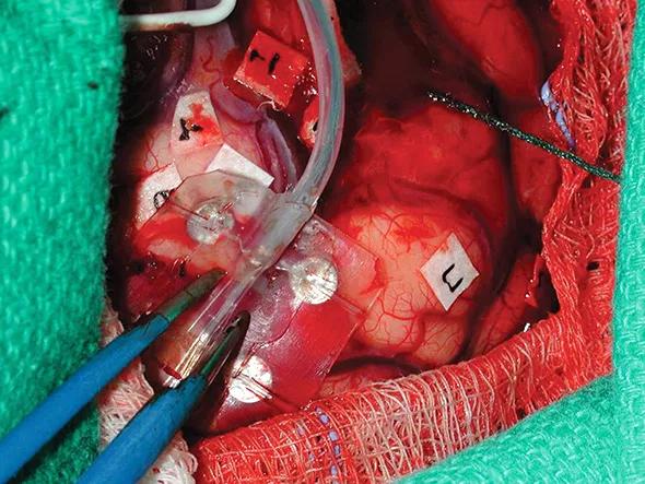
Image content: This image is available to view online.
View image online (https://assets.clevelandclinic.org/transform/5175d077-95db-4b93-9872-a08f27bf1108/figure1_jpg)
Figure 1. Intraoperative photograph of exposed brain showing placement of a grid (held by forceps) on the surface of the brain for electrocorticography. The number tags lie over the brain regions that control eloquent function, already mapped by cortical stimulation. The resection strategy can be developed in the operating room safely and cost-effectively.
Advertisement
Applying these techniques and creating an individualized plan for each TSC patient requires a team effort by experienced professionals. This is best done at a center with all facilities and personnel available under one roof.
At Cleveland Clinic’s Epilepsy Center, a multidisciplinary team of epilepsy professionals works together to perform a comprehensive multimodal evaluation of TSC patients and determine the best plan of care. A team of dedicated pediatric and adult epilepsy specialists, neurosurgeons, neuropsychologists, neuroradiologists, functional neuroimaging experts, and cognitive and behavioral experts meets at a weekly epilepsy management conference, together with health psychologists and bioethicists, to discuss the best individualized plan of care for patients with complex epilepsy.
Intricate decision-making needed
Patients with TSC often have to deal with involvement of other organs and systems, such as the kidneys, lungs, eyes, skin, heart and gastrointestinal system. This creates a need for intricate decision-making to deliver the most effective epilepsy care without harming the other organs or systems. That is why, at our center, an experienced epilepsy treatment team is embedded in an established multispecialty TSC program (Figure 2).
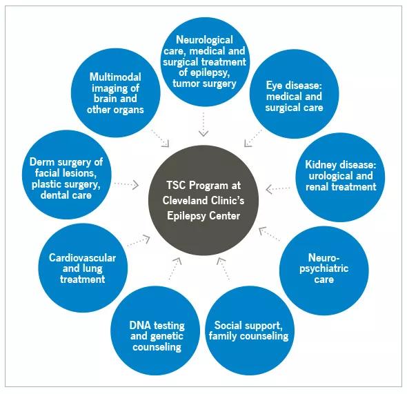
Image content: This image is available to view online.
View image online (https://assets.clevelandclinic.org/transform/51a00f40-cce0-4098-8610-068e312f8ccc/figure2_jpg)
Figure 2. Cleveland Clinic’s multispecialty tuberous sclerosis complex (TSC) program provides pediatric epilepsy care with collaborative support from multiple disciplines and resources under a single roof.
Dr. Gupta is Section Head of Pediatric Epilepsy in Cleveland Clinic’s Neurological Institute, where he established the TSC program in 2003.
Advertisement
Advertisement

New study advances understanding of patient-defined goals

Testing options and therapies are expanding for this poorly understood sleep disorder

Real-world claims data and tissue culture studies set the stage for randomized clinical testing

Digital subtraction angiography remains central to assessment of ‘benign’ PMSAH

Cleveland Clinic neuromuscular specialist shares insights on AI in his field and beyond

Findings challenge dogma that microglia are exclusively destructive regardless of location in brain

Neurology is especially well positioned for opportunities to enhance clinical care and medical training

New review distills insights from studies over the past decade