Locations:

Bringing OCT into the OR and more
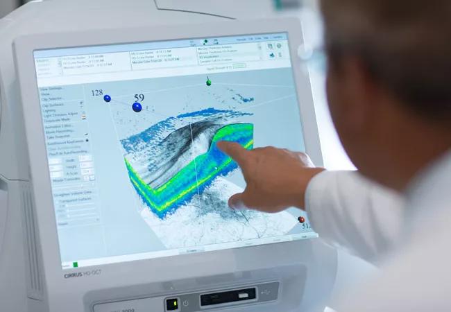
Image content: This image is available to view online.
View image online (https://assets.clevelandclinic.org/transform/0b0d882d-f21d-47e0-877a-78fca7dce831/16-EYE-2648-OTC-CQD-650p_jpg)
16-EYE-2648-OTC-CQD-650p
Advertisement
Cleveland Clinic is a non-profit academic medical center. Advertising on our site helps support our mission. We do not endorse non-Cleveland Clinic products or services. Policy
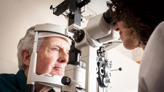
Noninvasive imaging has changed and continues to have the potential to change the way we practice. At Cole Eye Institute, we are continuing our tradition of pioneering new uses for optical coherence tomography (OCT).
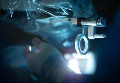
Today we are in the process of bringing OCT directly into our operating suites. Currently, when we treat macular holes or posterior vitreous retinal detachments in the clinic, we use OCT to visualize membranes, scar tissue and holes. But in the operating room, we have to guess.
Intraoperative OCT (iOCT) changes that. It tells us with 100 percent certainty whether or not we have achieved our surgical goals. We see the value of this advancement every time a patient comes for a second opinion after epiretinal membrane surgery at another facility. Using iOCT, we often spot remaining membrane that needs removal, invisible to a surgeon without iOCT.
Beyond simply moving a device from the clinic into the operating room, several members of our staff, including Justis P. Ehlers, MD, and Sunil K. Srivastava, MD, have been incorporating this technology into the way we perform surgery. Their innovative work actually places the OCT into the microscope. Our next step from here is to eliminate the traditional microscope altogether, incorporating OCT into a digital microscope that allows us to view an image on a large, high-resolution screen. Just like a camera’s images can be adjusted, we can modify a digital image to help us see, for example, the location of a tissue plane. This is extremely valuable in both surgery and in education.
Advertisement
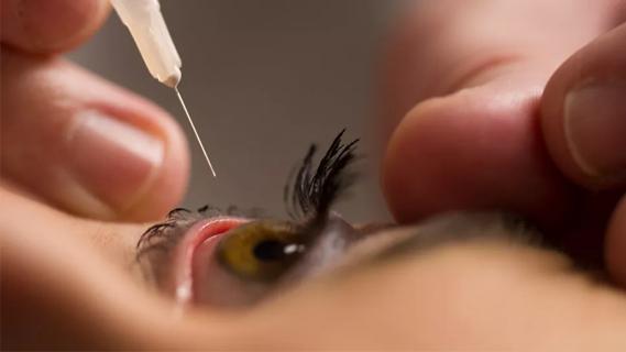
We also are working to use OCT angiography in place of indocyanine green angiography. Eliminating dye from the procedure is safer and faster, and yields higher quality images.
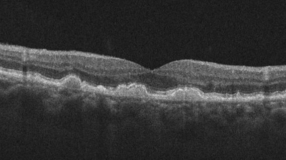
I’m also optimistic about our work developing the next generation of OCT, called swept source. Its longer wavelength penetrates deeper, giving us more information about the choroid and even the sclera than is possible with a traditional 850 mm laser. Swept source should allow us to see and perform 3D reconstructions and other techniques; it also lets us image the entire eye, which can be important to uveitis management. We hope to move our experimental swept source device into the clinic soon.
Cole Eye’s tradition of pushing the boundaries of ophthalmic imaging capabilities dates back to when David Huang, MD, PhD, one of the inventors of OCT, was on staff. Today, I am pleased to work with Drs. Srivastava and Ehlers, as well as Rishi Singh, MD, Sumit Sharma, MD, and others on pushing the envelope to find expanded applications for OCT.
Dr. Kaiser is the Chaney Family Endowed Chair of Ophthalmic Research and staff at Cole Eye Institute.
Advertisement
Advertisement
Early data shows risk is 73% higher in patients with lupus, 40% higher in patients with rheumatoid arthritis
Identifies weak spots in the cornea before shape change occurs
Study highlights the value of quantitative ultra-widefield angiography
Switching medications may decrease treatment burden and macular fluid

Interventions abound for active and stable phases of TED
Corneal imaging and interpretation play a major role
Cole Eye Institute imaging specialists are equal parts technician, artist and diagnostician

Effect of low-dose atropine and dual-focus contact lenses is unknown in patients with comorbid eye conditions