Much is possible when treating epilepsy in the developing brain
Advertisement
Cleveland Clinic is a non-profit academic medical center. Advertising on our site helps support our mission. We do not endorse non-Cleveland Clinic products or services. Policy
Kevin (a pseudonym) was brought by family to Cleveland Clinic at age 6.5 months for a third opinion on management of his seizure disorder, which first emerged at age 5 months in the setting of epidermal nevus syndrome. Other features of the condition included mild right hemihypertrophy, right linear alopecia, a midline facial nevus, linear hyperpigmentation on the right limbs and trunk, and mild coarctation of the aorta.
Six weeks before the visit, he developed seizures which escalated quickly to 10 to 100 epileptic spasms a day. His parents also noted that he was not moving his right arm and leg as much as the left. Since the most common brain malformation associated with epidermal nevus syndrome is hemimegalencephaly, a corresponding left hemispheric epilepsy syndrome was deemed most likely.
Video EEG revealed diffuse hypsarrhythmia with abundant spikes from multiple regions of both hemispheres, as well as nonlocalized EEG seizures during most of the spasms. However, the right posterior temporo-occipital region seemed disproportionately involved in the epileptogenic process. Interictal EEG often showed rhythmic periodic lateralized epileptiform discharge (PLED)-like runs of spikes and sharp waves from the right temporo-occipital region, and subclinical EEG seizures were also recorded from those areas. See Figure 1, which presents five EEGs in slideshow format.


Slide 1/5
MRI scan and PET imaging were available from age 5 months (see Figure 2, which presents four images in slideshow format).
Advertisement
The MRI showed the following on T2 sequences:
PET scan showed hypometabolism that was most pronounced in right temporo-occipital regions, with more intact and symmetrical metabolism in the frontal regions (Figure 2D).


Slide 1/4
At 6.5 months of age, he was being treated with phenobarbital and “medical” marijuana. He previously had been given oxcarbazepine and zonisamide without success.
Two therapy approaches were discussed by the Cleveland Clinic Epilepsy Center team and presented to the family, as follows:
Advertisement
The parents felt that epilepsy surgery was the best path. They had researched this extensively prior to consultation at Cleveland Clinic, and requested to proceed.
One of the following two courses could be taken:
Both centers where Kevin had been evaluated previously recommended hemispherectomy. The Cleveland Clinic team recommended the more conservative option, leaving open the possibility that a second procedure might be needed in the future.
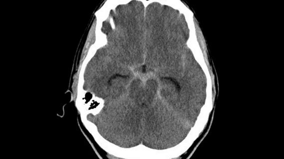
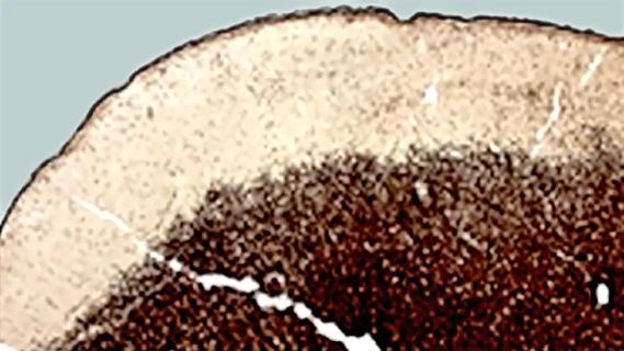
Kevin underwent a right temporo-occipital resection (Figure 3). Histology revealed cortical dysplasia, classified as International League Against Epilepsy (ILAE) type 1c. Findings included linear arrangements of neurons in the cortex and a loss of layer 2, with rare malpositioned neurons high in the cortex (Figure 4).

Figure 3

Figure 4
On days 1 to 4 following resection, Kevin had seizures in the form of subtle spasms. These abated spontaneously, and since then he has had no further seizures. After 16 months, EEG was normal and he was successfully weaned off all antiepileptic medications.
Advertisement
At his most recent appointment at 5.5 years of age, Kevin remained seizure-free, and his right hemi-neglect had resolved completely. He was developmentally appropriate and doing well in kindergarten.
Kevin’s case illustrates the following important issues in an infant with an underlying condition that is usually hemispheric.
Consider focality.It’s worth keeping in mind that even with a hemispheric neurological syndrome, the epilepsy process may be more localized. Clues to focality include:
Fixed hemiparesis and reversible hemi-neglect are difficult to distinguish in infancy. This fact makes motor-sparing surgery a favorable option when feasible, as it provides the opportunity for a functional motor deficit to resolve when the seizures are stopped.
In the immediate postoperative period, seizures may be self-limiting. Research has shown that patients may experience long-term seizure-free outcome despite some seizure recurrences in the first few weeks after surgery.
Early surgery may increase the chance for favorable seizure and developmental outcome. Had the family chosen trying the medical option first, we would have strongly encouraged them to not let the trial last more than a few months without success. There is some evidence to indicate that the longer epilepsy is left uncontrolled, the more likely it is to become fixed — and unfixable. Another reason to consider early surgery is plasticity in the developing brain, which affords opportunity for improved neurological outcome.
Advertisement
In this case, an excellent outcome resulted from a controversial decision to choose a more conservative surgical approach. It is important not to forget what’s possible when dealing with epilepsy in the developing brain.
Dr. Wyllie, Professor of Neurology at Cleveland Clinic Lerner College of Medicine, is a pediatric epilepsy specialist at Cleveland Clinic.
Advertisement

New study advances understanding of patient-defined goals

Testing options and therapies are expanding for this poorly understood sleep disorder

Real-world claims data and tissue culture studies set the stage for randomized clinical testing

Digital subtraction angiography remains central to assessment of ‘benign’ PMSAH

Cleveland Clinic neuromuscular specialist shares insights on AI in his field and beyond

Findings challenge dogma that microglia are exclusively destructive regardless of location in brain

Neurology is especially well positioned for opportunities to enhance clinical care and medical training

New review distills insights from studies over the past decade