Locations:

An underrecognized but significant complication in pediatric patients
Image content: This image is available to view online.
View image online (https://assets.clevelandclinic.org/transform/abc727e2-db9a-4ce9-9bbb-b729695d3908/18-NEU-782-Hero_650x450_jpg)
18-NEU-782-Hero_650x450
By Neil Friedman, MBChB; Manikum Moodley, MD; and A. David Rothner, MD
Advertisement
Cleveland Clinic is a non-profit academic medical center. Advertising on our site helps support our mission. We do not endorse non-Cleveland Clinic products or services. Policy
Neurofibromatosis type 1 (NF1) is the most common of the neurocutaneous syndromes, affecting about 1 in 3,000 individuals — or at least 1 million individuals worldwide. First described as a nosological entity in 1882, it affects both sexes and all races and ethnic groups.
Approximately half of cases arise as new, spontaneous mutations. Most people with NF1 have symptoms by age 10. As with many autosomal dominant disorders, the phenotypic expression is highly variable. The risk of the condition’s various manifestations changes across developmental stages and can vary significantly between individuals and even within a family, making prognostication difficult. While NF1 is not a life-threatening condition for most individuals, its two most feared complications — and potentially life-threatening associations — are malignancy and vascular dysplasias.
NF1 vasculopathy is an uncommon but significant and underrecognized complication of neurofibromatosis. While vascular abnormalities in NF1 have long been recognized,1 their precise incidence is unknown and has not been evaluated in prospective studies.
The pathophysiology also remains incompletely understood. Deficiency of neurofibromin, the protein product of the NF1 gene, in the vessel endothelial and smooth muscle cells is the likely pathogenesis of NF1-associated vasculopathy.2 It is thought to lead to excessive proliferation of the vascular endothelial and/or smooth muscle cells. Neurofibromin is also important in maintaining integrity of the endothelial cell layer, and its mutation alters this integrity, leading to unopposed proliferation of the vascular smooth muscle cells. Why only a single artery or a few selective arteries are affected in a particular individual remains unclear. No genotype-phenotype correlation has been shown.
Advertisement
Stenosis or occlusion of vessels may result in cerebral or visceral infarcts. Other complications include aneurysms, with risk of rupture and hemorrhage, and arteriovenous fistulae. While the entire vascular tree may be affected, more commonly the renal, celiac, iliac, abdominal aorta and cerebral vasculature are implicated.
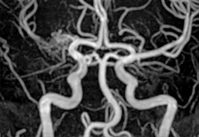
Image content: This image is available to view online.
View image online (https://assets.clevelandclinic.org/transform/c4e13d66-0584-4dc5-a5b9-3c17424a9706/18-NEU-782-Inset_650x450_jpg)
Figure. Unilateral moyamoya in NF1 vasculopathy: MRA of the brain showing occlusion of the right M1 segment of the middle cerebral artery with reconstitution of the M2 segment secondary to a tangle of collateral vessels. Reprinted from Ghosh et al.3 © The Authors 2013.
Cerebrovascular manifestations include stenoses, occlusions, ectasia, fusiform aneurysm formation and arteriovenous fistulae involving the anterior and/or posterior circulation. Moyamoya vasculopathy is most commonly seen and is often unilateral, typically involving the anterior circulation in children (Figure).3 The extracranial carotid and vertebral arteries may also be involved. Prevalence of cerebrovascular manifestations in NF1 is estimated at 2.5 percent to 6 percent.3 Although most patients with cerebral vasculopathy are asymptomatic,3,4 clinical and radiologic worsening often occurs with risk of transient ischemic events and stroke.4,5
A number of questions remain regarding the nature of the cerebral vasculopathy in NF1. Given that the natural history, disease progression and prognosis have not been well studied, optimal monitoring, imaging and treatment guidelines have not been well established. Controversy exists regarding routine vascular screening in NF1. There is also no consensus regarding indication and timing of revascularization surgery versus medical management with antiplatelet therapy in asymptomatic individuals with moyamoya or other cerebral vasculopathies.
Advertisement
The neurovascular and cardiovascular complications of neurofibromatosis have long been an area of focus and interest for Cleveland Clinic’s multidisciplinary, comprehensive neurofibromatosis clinic, which is a member of the Neurofibromatosis Clinic Network established by the Children’s Tumor Foundation. Our clinic sees 100 or more patients with NF1 annually, at least 80 percent of them under age 18.
Given the infrequent occurrence of cerebral vasculopathy in NF1, however, progress will certainly depend on more than single-center efforts. We look forward to the prospect of multicenter studies to answer many of the unanswered questions above and develop guidelines for the medical and/or surgical management of NF1 vasculopathy.
Advertisement
Dr. Friedman (friedmn@ccf.org) is Director of Cleveland Clinic’s Center for Pediatric Neurosciences, and Drs. Moodley and Rothner are pediatric neurologists within the center.
Advertisement
Advertisement

New study advances understanding of patient-defined goals

Testing options and therapies are expanding for this poorly understood sleep disorder

Real-world claims data and tissue culture studies set the stage for randomized clinical testing

Digital subtraction angiography remains central to assessment of ‘benign’ PMSAH

Cleveland Clinic neuromuscular specialist shares insights on AI in his field and beyond

Findings challenge dogma that microglia are exclusively destructive regardless of location in brain

Neurology is especially well positioned for opportunities to enhance clinical care and medical training

New review distills insights from studies over the past decade