Locations:

Cell-specific response to viral infection identified

Image content: This image is available to view online.
View image online (https://assets.clevelandclinic.org/transform/4ed9006f-60b3-47c7-8a66-d08be745f8ae/15-EYE-2742-SearsResearch-CQD-650x450_jpg)
15-EYE-2742-SearsResearch-CQD-650×450
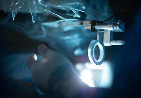
Researchers at Cleveland Clinic are shedding light on biological processes involved in glaucoma — specifically how cells choose between inflammation and apoptosis, or programmed cell death. These insights could eventually advance the search for a neuroprotective treatment that might slow the course of the disease.
Advertisement
Cleveland Clinic is a non-profit academic medical center. Advertising on our site helps support our mission. We do not endorse non-Cleveland Clinic products or services. Policy
“Recent biochemical and histological studies have shown that glaucomatous ganglion cell death is a form of slow apoptosis. One possibility is that glaucomatous atrophy may involve ganglion cell inflammation prior to apoptosis,” says Nathaniel Sears, MD, a researcher at Cleveland Clinic’s Cole Eye Institute.
Dr. Sears notes that studies using electroretinography and visual field testing have shown that in patients first diagnosed with glaucoma, some ganglion cells are already dead, while others are dysfunctional but still viable — as if caught in a chronic inflammatory state. “These conclusions are largely based on the partial recovery of retinal function once IOP is lowered,” he says.
“Ganglion cell apoptosis may be a protracted process that depends on equilibrium between an inflamed, pre-apoptotic state and overt apoptosis,” Dr. Sears says. “A neuroprotectant that could be used to retard the death of involved neurons might increase the chance for recovery, or at least slow the course of glaucoma, especially after the patient’s intraocular pressure (IOP) is lowered,” he says.
Dr. Sears has investigated the hypothesis that there is a regulatory switch between inflammation and apoptosis. As a model for these investigations, he has studied the pathway by which cells sense viral infection via the receptor retinoic acid-inducible gene I (RIG-I). “When activated, RIG-I initiates the phosphorylation of interferon response factor 3 (IRF3), leading to the Type-1 interferon antiviral response,” he says.
Advertisement
As a medical student, Dr. Sears found that after activation and initiation of a Type 1 interferon response, IRF3 degrades to a separate but identifiable fragment that maintains immunoreactivity — similar to the mature protein.
“Pursuing a hunch that this was a fragment of IRF3 and part of the regulation of IRF3 activity, I showed that full length IRF3 has a caspase recognition sequence. I then definitively demonstrated that caspase-8 cleaves IRF3 after activation, targeting it for degradation to inhibit downstream gene induction,” he says.
Since caspase-8 activation leads to apoptosis, its interaction with the IRF3 substrate is a form of molecular rheostat that determines whether a cell’s fate is inflammation or apoptosis, according to Dr. Sears.
“We are currently working with several ocular cell lines to characterize their specific response to identical viral stimulus,” he says. “Although it is too soon to draw conclusions, I have thus far determined that differences in cellular mortality coincide with specific markers of IRF3 activation,” he says.
Dr. Sears and colleagues have correlated increased cell death to increased expression of IFIT, ordinarily an IRF3-regulated gene. The researchers have shown that overexpression of the IRF3 fragment can initiate cell death. Their preliminary studies indicate that IRF3-independent IFIT expression is a biomarker of cell death. “We’re now developing a model in ganglion cells to see if it is possible to regulate their apoptotic switch,” he adds.
Advertisement
“Glaucoma is a disease that a patient will have to cope with for life,” Dr. Sears says. Once ganglion cells die, blindness is irreversible in these patients. So, ultimately, neuroprotection may be able to inhibit susceptible retinal ganglion cells from choosing death after IOP reduction — “to prevent the relentless progression of blindness in glaucoma,” he says.
Advertisement
Advertisement
Early data shows risk is 73% higher in patients with lupus, 40% higher in patients with rheumatoid arthritis
Identifies weak spots in the cornea before shape change occurs
Study highlights the value of quantitative ultra-widefield angiography
Switching medications may decrease treatment burden and macular fluid

Interventions abound for active and stable phases of TED
Corneal imaging and interpretation play a major role
Cole Eye Institute imaging specialists are equal parts technician, artist and diagnostician

Effect of low-dose atropine and dual-focus contact lenses is unknown in patients with comorbid eye conditions