Locations:

Examination and echocardiography play a critical role in diagnosis
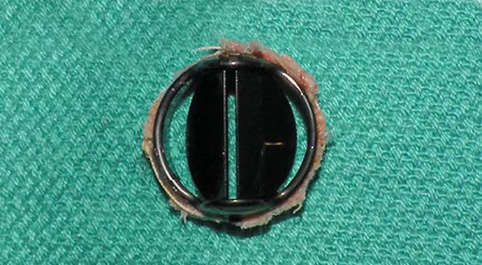
Image content: This image is available to view online.
View image online (https://assets.clevelandclinic.org/transform/57d7906e-4bf2-4a0a-8b8f-469f6c9fceb0/Surgical_excision_690x380_jpg)
Surgical_excision_690x380
Newton Wiggins, MD
Cardiology Fellow, Cleveland Clinic
Advertisement
Cleveland Clinic is a non-profit academic medical center. Advertising on our site helps support our mission. We do not endorse non-Cleveland Clinic products or services. Policy
A 60-year-old woman presented to the Cleveland Clinic outpatient department for evaluation of progressive shortness of breath.
She was diagnosed with rheumatic heart disease in 1977 during her second pregnancy. She underwent a mitral commissurotomy for treatment of mitral stenosis in 1978. In 1983, her mitral valve was replaced with a porcine valve. In 1993, her aortic valve and porcine mitral valve were replaced with St. Jude mechanical valves. Finally, in 1997, she had her mechanical aortic valve (#19) replaced with another St. Jude mechanical aortic valve (#21). During this surgery, she also underwent placement of an ascending aortic patch graft.
She then did well for approximately 15 years. For the past two years, she has struggled with progressive shortness of breath, fatigue, and exercise intolerance despite medication and dietary compliance. After a local echocardiogram found an increased transprosthetic gradient across her aortic valve, she was referred to the Cleveland Clinic for further evaluation.
Her vital signs in clinic were normal. She had a normal body mass index. She appeared well and was able to move around the examination room without difficulty. There was no jugular venous distention with the patient reclined at 45 degrees. Pulmonary examination was normal. She had a regular rhythm and a nondisplaced apical impulse. Auscultation of her precordium revealed mechanical first and second heart sounds. A loud systolic murmur was heard best at the upper sternal borders and varied in intensity over time. There was no correlation with the respiratory cycle. Peripheral edema was not present. The rest of her examination was normal.
Advertisement
Both transthoracic and transesophageal echocardiography showed that the mechanical aortic valve was stenotic, but the severity of the stenosis was difficult to determine due to shielding from the mechanical mitral valve, which was functioning normally.
She was then sent for cinefluoroscopy of the mechanical aortic valve, which showed that the prosthetic leaflet closest to the right coronary sinus was intermittently fixed in the closed position. This malfunction was thought to be secondary to pannus formation, and she was referred for cardiac surgery.
Approximately one month later, she successfully underwent fourth time redo median sternotomy with re-replacement of her aortic valve as well as coronary artery bypass grafting for one-vessel coronary artery disease. Her mechanical aortic prosthesis was found to have pannus formation both above and below the valve, confirming the mechanism of her intermittent obstruction.
She was discharged home approximately one week after surgery and was doing well at her initial telephone follow-up visit.
The manifestations of prosthetic valve dysfunction vary depending on the valve type, valve location, and the clinical factors of the patient. One such manifestation is valve obstruction, which can present with a spectrum of symptoms ranging from an incidental finding to complete cardiovascular collapse. The causes of obstruction include pannus, thrombus, and vegetation formation.
As in this patient, examination often plays a critical role in the diagnosis especially when there is a change in the character of mechanical heart sounds. Echocardiography also plays a key role in the evaluation usually by identifying an increased transprosthetic gradient. Quantitative evaluation of leaflet motion by echocardiography may be accurately achieved for patients with mechanical valves. However, in patients with both mechanical mitral and aortic valves, shielding can limit visualization particularly of the aortic valve. Cinefluoroscopy remains a very useful and “gold-standard” technique for evaluation of leaflet motion.(1)
Advertisement
Treatment depends on the mechanism of the obstruction but usually involves surgery, especially when pannus formation is the cause. Thrombosis can sometimes be treated with fibrinolysis in certain patients after careful assessment of the risks and benefits.(2)
Prosthetic valve dysfunction can manifest in a variety of ways and as highlighted in this case study, sometimes requires a high index of suspicion to make the diagnosis. Intermittent fixation of a prosthetic disc is uncommon but can represent an atypical consequence of pannus formation. This diagnosis can easily be missed without an adequate duration of auscultation or imaging. This case also underscores the utility of cinefluoroscopy in the assessment of mechanical valves particularly if inadequately visualized by echocardiography.
References
1. Muratori, M et al. Feasibility and diagnostic accuracy of quantitative assessment of mechanical prostheses leaflet motion by transthoracic and transesophageal echocardiography in suspected prosthetic valve dysfunction. Am J Cardiol 2006;97:94-100.
2. Nushimura, R et al. 2014 ACC/AHA guideline for the management of patients with valvular heart disease. Circulation 2014;129:e521-e643.
Advertisement
Advertisement

A sampling of outcome and volume data from our Heart & Vascular Institute

Concomitant AF ablation and LAA occlusion strongly endorsed during elective heart surgery

Large retrospective study supports its addition to BAV repair toolbox at expert centers

Young age, solid tumor, high uptake on PET and KRAS mutation signal risk, suggest need for lobectomy

Surprise findings argue for caution about testosterone use in men at risk for fracture

Residual AR related to severe preoperative AR increases risk of progression, need for reoperation

Findings support emphasis on markers of frailty related to, but not dependent on, age

Provides option for patients previously deemed anatomically unsuitable