Locations:

Total ankle replacement yields big improvements
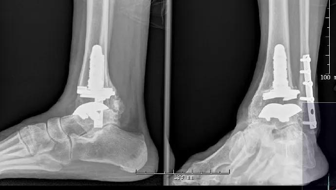
Image content: This image is available to view online.
View image online (https://assets.clevelandclinic.org/transform/3dcc87f0-59df-4a42-b435-626f88ecc42d/Sferra-Hero-Image-690x380pxl_jpg)
Sferra-Hero-Image-690x380pxl
A 64-year-old woman presented for evaluation of left ankle pain. She had sustained a left bimalleolar ankle fracture 12 years previously, which was treated with operative reduction and internal fixation at another facility. On presentation, she reported a history of several years of ankle pain with ambulation. Previous treatments included steroid injections, NSAIDs and Arizona-type AFO bracing.
Advertisement
Cleveland Clinic is a non-profit academic medical center. Advertising on our site helps support our mission. We do not endorse non-Cleveland Clinic products or services. Policy
On physical exam, she had a BMI of 29 and a severely antalgic gait. She had very limited motion in her ankle, which was exquisitely tender to palpation at the tibiotalar joint. Subtalar motion and midfoot motion were supple and nontender. Anteroposterior (AP), lateral and oblique weight-bearing X-rays (Figure 1) demonstrated severe left tibiotalar degenerative joint disease. Her subtalar and transverse tarsal joints were well maintained. There was retained hardware in the distal fibula and tibia.

Image content: This image is available to view online.
View image online (https://assets.clevelandclinic.org/transform/efe69a7c-0590-4634-bdba-8d389b04c00b/Sferra-Figure-1-Inset-590pxl-width_jpg)
Figure 1. Preoperative weight-bearing AP and lateral X-rays demonstrating severe osteoarthritis. No deformity is present, and adjacent joints have been spared.
She underwent total ankle replacement with the Inbone Total Ankle System and removal of her medial hardware. Intraoperatively, she was found to have a chronically ruptured tibialis anterior tendon. The remnant of this tendon was tenodesed to the extensor hallucis longus tendon.
She remained non-weight-bearing in a cast for three weeks. Afterward, she was transitioned to a non-weight-bearing pneumatic walking boot during the day to begin gentle range-of-motion exercises three times daily. At postoperative week 8, her weight-bearing was increased by 25 percent every four to five days in the boot.
At her last follow-up, 14 months after surgery, she had no complaints and reported no pain. She did have some active ankle dorsiflexion, which on clinical exam appeared to be through her tibialis anterior. Postoperative X-rays at that time revealed well-fixed components (Figure 2).
Advertisement

Image content: This image is available to view online.
View image online (https://assets.clevelandclinic.org/transform/e9fa125b-1ab0-47d3-bff3-0af012c28f09/Sferra-Figure-2-Inset-590pxl-width_jpg)
Figure 2. Postoperative weight-bearing AP and lateral oblique X-rays demonstrating the Inbone TAR implant.
Advertisement
Advertisement

Biologic approaches, growing implants and more
Study reports zero infections in nearly 300 patients
How to diagnose and treat crystalline arthropathy after knee replacement
Study finds that fracture and infection are rare
Center will coordinate, interpret and archive imaging data for all multicenter trials conducted by the foundation’s Osteoarthritis Clinical Trial Network

Reduced narcotic use is the latest on the list of robotic surgery advantages
Cleveland Clinic specialists offer annual refresher on upper extremity fundamentals
Cleveland Clinic orthopaedic surgeons share their best tips, most challenging cases and biggest misperceptions