Locations:

Identifying the cause of groin pain, numbness in runner
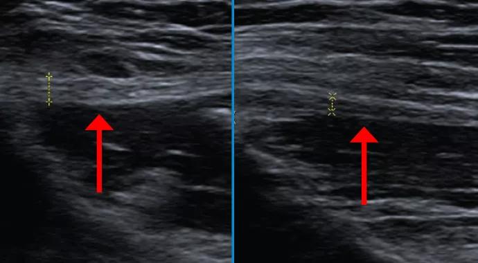
Image content: This image is available to view online.
View image online (https://assets.clevelandclinic.org/transform/16b07b3c-cbf7-4b2f-8970-24efea90346a/14-ORT-1685-Polster-Hero-Image-690x380pxl_jpg)
14-ORT-1685-Polster-Hero-Image-690x380pxl
Advertisement
Cleveland Clinic is a non-profit academic medical center. Advertising on our site helps support our mission. We do not endorse non-Cleveland Clinic products or services. Policy
A 34-year-old woman with no significant medical history presented with a chief complaint of left groin pain that came on suddenly one year earlier during a 15-mile marathon-training run. She reported that the pain was aggravated by most activities. She also had intermittent left lateral thigh numbness.
She had a prior workup at an outside institution that included lumbar spine MRI, diagnostic laparoscopy to evaluate for ovarian pathology, colonoscopy and cystoscopy, all of which were negative. Physical exam demonstrated localized tenderness about 2 cm medial and inferior to the left anteriorsuperior iliac spine. Strength was normal.
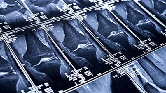
Cleveland Clinic sports health orthopaedic surgeon James Rosneck, MD, referred the patient for dedicated ultrasound neurography, which demonstrated asymmetric thickening of the left femoral cutaneous nerve just distal to the left anterior-superior iliac spine, where there was associated tenderness. Figure 1 contrasts a longitudinal view of the abnormally thickened lateral femoral cutaneous nerve (left image) with a longitudinal view of the contralateral normal nerve (right image). Figure 2 demonstrates the same side-to-side difference in the transverse plane.
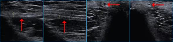
Image content: This image is available to view online.
View image online (https://assets.clevelandclinic.org/transform/0a4246d3-0256-411b-ad83-bb06e04615f7/14-ORT-1685-Polster-Inset-Image-590pxl-width_jpg)
Figure 1. Longitudinal views on ultrasound neurography showing an abnormally thickened lateral femoral cutaneous nerve (arrow, left image) in contrast with the contralateral normal nerve (arrow, right image).
Figure 2. Transverse views on ultrasound neurography demonstrating the same side-to-side difference as in Figure 1.
Advertisement
Based on these imaging findings and the clinical history, the patient was diagnosed with lateral femoral cutaneous neuropathy. She was relieved to have the likely cause of her symptoms found after numerous other normal studies.
Ultrasound neurography is the specialized use of ultrasound imaging to evaluate peripheral nerves. This technique offers several potential advantages relative to MR neurography, including:
Ultrasound neurography requires high-frequency ultrasound probes and detailed knowledge of peripheral nerve anatomy. For these reasons, ultrasound neurography is performed at Cleveland Clinic by subspecialty-trained musculoskeletal radiologists and neurologists.
Dr. Polster is a musculoskeletal radiologist in the Department of Diagnostic Radiology.
Advertisement
Advertisement

Biologic approaches, growing implants and more
Study reports zero infections in nearly 300 patients
How to diagnose and treat crystalline arthropathy after knee replacement
Study finds that fracture and infection are rare
Center will coordinate, interpret and archive imaging data for all multicenter trials conducted by the foundation’s Osteoarthritis Clinical Trial Network

Reduced narcotic use is the latest on the list of robotic surgery advantages
Cleveland Clinic specialists offer annual refresher on upper extremity fundamentals
Cleveland Clinic orthopaedic surgeons share their best tips, most challenging cases and biggest misperceptions