Locations:

Optimizing quality of cartilage-forming cells
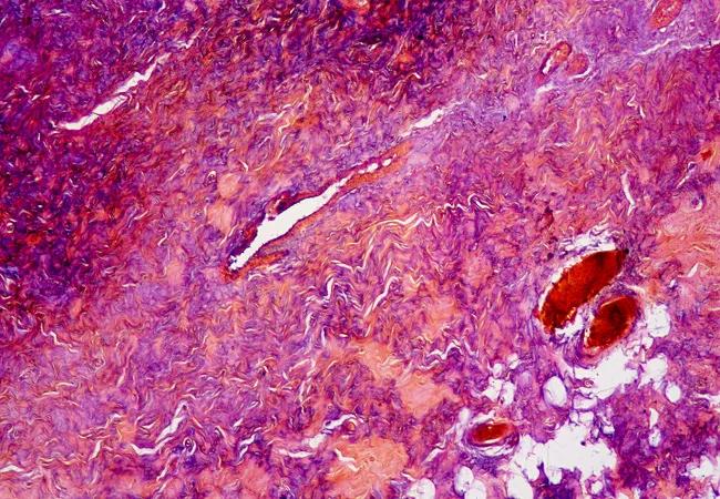
Image content: This image is available to view online.
View image online (https://assets.clevelandclinic.org/transform/20e3476d-65ea-4478-b97f-b162793621e2/17-ORT-1163-Mantripragada-Hero-Image-650x450pxl_jpg)
17-ORT-1163-Mantripragada-Hero-Image-650x450pxl
By Venkata P. Mantripragada, PhD, and George F. Muschler, MD
Advertisement
Cleveland Clinic is a non-profit academic medical center. Advertising on our site helps support our mission. We do not endorse non-Cleveland Clinic products or services. Policy
How to repair, replace or regenerate cartilage has been the Holy Grail for orthopaedic researchers worldwide for more than five decades. Great strides have been made, and orthopaedic surgeons and researchers have developed numerous strategies to attempt to form new cartilage in the knee, including microfracture, periosteal flaps, cartilage transplantation and autologous chondrocyte implantation.
The most widely used cell therapy procedure, using bone marrow aspirate concentrate (BMAC) for the treatment of knee osteoarthritis, is yet another manifestation of the high value we and our patients place on cartilage health, preservation and restoration.
Each strategy has something in common: the need for a source of cells that can form new cartilage. These chondrogenic connective tissue progenitor cells (CTP-Cs) differ from CTPs that tend to form other tissues (e.g., bone, fat or scar).
Unfortunately, none of the repair options developed to date has proven 100 percent successful in restoring the native articular cartilage structure with hyaline cartilage. To determine the best strategy and to have a profound effect on the outcome, we are trying to answer a number of specific questions about the nature of the cells and their biological performance:
Advertisement
Our research team, from the departments of Orthopaedic Surgery and Biomedical Engineering, is diligently working to answer these questions. We collect discarded samples of cartilage, synovium, periosteum and retropatellar fat from knees of patients undergoing elective total knee arthroplasty (those who grant permission). Most patients also graciously agree to allow us to collect bone marrow by aspiration from the iliac crest. These samples provide an exceptional opportunity to directly compare the quality of CTP-C tissue sources. Our goal is to measure both the number of CTPs and their relative ability to form cartilage (i.e., develop an assay of CTP-Cs).
In order to perform our analysis of cultured CTP-Cs in a reliable and reproducible manner, we use a customized robot to collect the high-resolution images needed to measure and characterize CTPs. This robot was designed and built in the Muschler laboratory at Cleveland Clinic in collaboration with Parker Hannifin Corp., a Cleveland company that focuses on motion and control technologies. Automated image analysis software was also developed in the laboratory to extract detailed quantitative information from each CTP-C colony, based on cell surface markers and extracellular matrix molecules (Figures 1 and 2). These methods have become valuable well beyond our laboratory and have been incorporated into the ASTM Standard Methods for Automated Cell and Colony Analysis.1

Image content: This image is available to view online.
View image online (https://assets.clevelandclinic.org/transform/af1d5c73-abe6-40d9-a270-a00ff687b9ef/17-ORT-1163-Mantripragada-Inset-Image-01_jpg)
Figure 1. Analysis of two colonies derived from CTPs resident in A. synovium and B. cartilage. CTPs from synovium proliferate more rapidly and form larger colonies, which are less dense. In contrast, CTPs from cartilage form smaller and more densely packed colonies in both 2-D and 3-D cultures.
Advertisement

Image content: This image is available to view online.
View image online (https://assets.clevelandclinic.org/transform/a6b3891b-9c44-4cc8-bf52-e7b12a71d73d/17-ORT-1163-Mantripragada-Inset-Image-02_jpg)
Figure 2. Time-lapse videomicroscopy allows quantitative analysis of colony formation, starting with the colony founding CTP-C (left panels). The colonies formed by two founding cells are presented using phase-contrast microscopy (middle panels) and quantitative three-color immunofluorescence (right panels). Large variation is seen in colony morphology. The upper colony shows tightly packed cuboidal cells with chondrogenic morphology. The lower colony comprises loosely packed, elongated fibroblastic cells at day 12.
In our current study, tissue sources from 20 patients were compared with respect to cell concentration (cells per gram of tissue), prevalence (CTPs per million cells plated) and CTP concentration (CTPs per gram of tissue). The table, below, shows the comparison between four tissue sources. Periosteum and synovium had a high cell concentration, but the prevalence of CTPs was low. Fat, on the other hand, was less cellular, but had a relatively higher prevalence of CTPs. Cartilage performed the best in terms of concentration, prevalence, expression of chondrogenic markers of CTP-Cs and the ability to grow in 3-D gels.

Image content: This image is available to view online.
View image online (https://assets.clevelandclinic.org/transform/076ddcfb-4f66-4f47-ad19-c62477fb2405/17-ORT-1163-Mantripragada-Inset-Image-03_jpg)
The rational development of cell therapy based on adult progenitor cells will require a qualitative and quantitative assessment to define optimal sources of cells for cartilage tissue repair. This source should have several characteristics:
Advertisement
Further work in identifying optimal sources of CTP-Cs will be essential for the ongoing development of safe, efficient and reliable therapies for the repair and regeneration of cartilage and joint preservation.
This work is supported by a National Institutes of Health R01 grant, AR063733.
Reference
Dr. Mantripragada is a postdoctoral fellow in the Biomedical Engineering Department at Cleveland Clinic’s Lerner Research Institute. Dr. Muschler is a professor of orthopaedic surgery, specializing in all aspects of knee and hip replacements. He is Director of the Regenerative Medicine Laboratory at Cleveland Clinic, where he conducts research on bone and cartilage tissue regeneration.
Advertisement
Advertisement

Biologic approaches, growing implants and more
Study reports zero infections in nearly 300 patients
How to diagnose and treat crystalline arthropathy after knee replacement
Study finds that fracture and infection are rare
Center will coordinate, interpret and archive imaging data for all multicenter trials conducted by the foundation’s Osteoarthritis Clinical Trial Network
Reduced narcotic use is the latest on the list of robotic surgery advantages
Cleveland Clinic specialists offer annual refresher on upper extremity fundamentals
Cleveland Clinic orthopaedic surgeons share their best tips, most challenging cases and biggest misperceptions