Locations:

Better treatment urgently needed
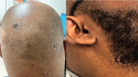
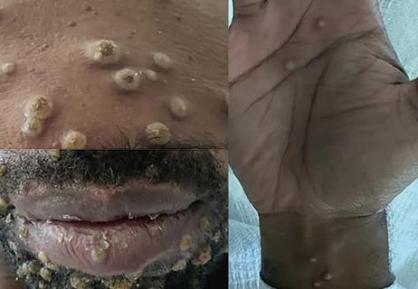
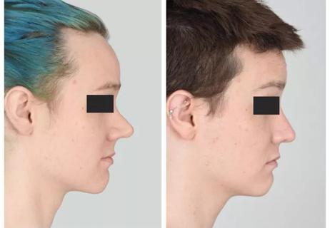
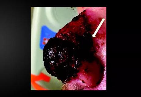
Dystrophic calcinosis is the most common form of calcinosis, the deposition of insoluble calcium salts in various tissues. It occurs most frequently in patients with underlying autoimmune rheumatic diseases such as scleroderma and dermatomyositis.
Advertisement
Cleveland Clinic is a non-profit academic medical center. Advertising on our site helps support our mission. We do not endorse non-Cleveland Clinic products or services. Policy
These slides depict calcinosis in several patients, including a 29-year-old female with juvenile dermatomyositis since age 7 whose myositis has remained in remission on prednisone, hydroxychloroquine and intravenous immunoglobulin (IVIG) infusions. However, her subcutaneous calcium deposits progressed relentlessly, ultimately covering her entire body like an exoskeleton.
These cases demonstrate the devastating nature of dystrophic calcinosis and the urgent need for further research to develop effective therapies.

Image content: This image is available to view online.
View image online (https://assets.clevelandclinic.org/transform/1ac47ae7-51a8-4f8c-b193-ddd34d6cb551/800x550-Chatterjee-Slide-07-150x103_jpg)

Image content: This image is available to view online.
View image online (https://assets.clevelandclinic.org/transform/321ff46f-2a72-4a46-8836-2e6b77eb87b7/800x550-Chatterjee-Slide-09-150x103_jpg)

Image content: This image is available to view online.
View image online (https://assets.clevelandclinic.org/transform/93a789b6-0d3c-4646-80f8-f8bd245285e5/800x550-Chatterjee-Slide-02-150x103_jpg)

Image content: This image is available to view online.
View image online (https://assets.clevelandclinic.org/transform/ae084dd7-5351-4ca8-98e2-8dfbce89a7e4/800x550-Chatterjee-Slide-05-150x103_jpg)
Advertisement

Image content: This image is available to view online.
View image online (https://assets.clevelandclinic.org/transform/37e586b7-5943-4def-8d41-4eb211d1447a/800x550-Chatterjee-Slide-01-150x103_jpg)

Image content: This image is available to view online.
View image online (https://assets.clevelandclinic.org/transform/6cc9d8ae-c70e-4855-83e0-a4e9574e2b4c/800x550-Chatterjee-Slide-06-150x103_jpg)

Image content: This image is available to view online.
View image online (https://assets.clevelandclinic.org/transform/78e56720-7fb4-4e3d-8035-5c652dd49a69/800x550-Chatterjee-Slide-03-150x103_jpg)

Image content: This image is available to view online.
View image online (https://assets.clevelandclinic.org/transform/e0f6cc56-b0f7-4a5f-be24-94fc3f5460ba/800x550-Chatterjee-Slide-04-150x103_jpg)

Image content: This image is available to view online.
View image online (https://assets.clevelandclinic.org/transform/879f8391-8a37-4762-b058-b29689eaeb16/800x550-Chatterjee-Slide-08-150x103_jpg)
Slide 1/9
Images and text provided by Soumya Chatterjee, MD, Director, Scleroderma Program, Department of Rheumatic and Immunologic Diseases.
Advertisement
Advertisement

Here is the awesome subtitle.

Consider secondary syphilis in the differential of annular lesions
Persistent rectal pain leads to diffuse pustules
Techniques are borrowed from rhinoplasty, malar augmentation and others

Two cases — both tremendously different in their level of complexity — illustrate the core principles of nasal reconstruction
Stress and immunosuppression can trigger reactivation of latent virus

Low-dose, monitored prescription therapy demonstrates success