Locations:

Insights from 80 paired comparisons of 3T vs. 7T images
Image content: This image is available to view online.
View image online (https://assets.clevelandclinic.org/transform/d78d6ca6-a3db-42ed-8143-7dfa7c79d904/16-NEU-1938-Jones-650x450-1_jpg)
16-neu-1938-jones-650×450
Advertisement
Cleveland Clinic is a non-profit academic medical center. Advertising on our site helps support our mission. We do not endorse non-Cleveland Clinic products or services. Policy
An advanced 7-tesla (7T) MRI scanner was installed at Cleveland Clinic in 2013 and has now been in active use for more than two years. The principal advantage of 7T MRI over MRI scanning at lower magnetic field strengths is increased signal, which can provide smoother images (higher signal-to-noise ratio), faster scanning and higher resolution. The latter is the factor most likely to expand the future clinical application of 7T MRI.
RELATED: The Art of 7T Imaging (Slideshow)
In addition to research and development studies, Cleveland Clinic’s 7T scanner has been used, with IRB approval, to scan patients with neurological disease for the explicit purpose of comparing lesion conspicuity between 7T images and images obtained previously at lower magnetic field strengths.
To date, 134 patients have undergone this type of comparative imaging, with diseases including epilepsy, multiple sclerosis, amyotrophic lateral sclerosis, traumatic brain injury, orbital neoplasm, vasculitis, brain tumors and others. Research is now underway evaluating the clinical utility of 7T MRI for enhancing the diagnosis of epilepsy, with preliminary results showing that 7T images enhance previous findings in nearly half of patients imaged.
To define 7T’s incremental contribution to image quality, 11 members of Cleveland Clinic’s neuroradiology staff assessed 80 paired images of various lesions — one at 7T and one at 3T — in a blinded manner. Each image was scored on a five-point scale for lesion conspicuity (clearly superior, mildly superior, equal, mildly inferior, clearly inferior).
Advertisement
Across the full set of image pairs, the overall assessment of the neuroradiologists was that lesion conspicuity at 7T was mildly superior to lesion conspicuity at 3T, averaged over the large assortment of different diseases and sequences. Depending on the sequence and disease, many specific examples were considered clearly superior by nearly all neuroradiologists. The figure below presents four of the image pairs as an example.
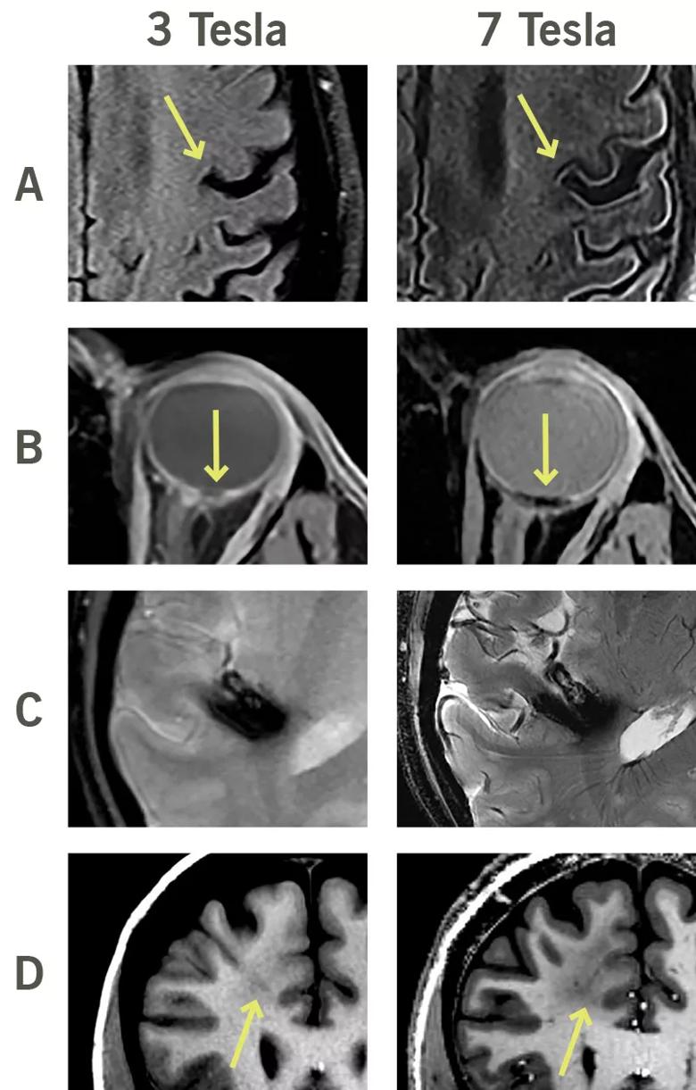
Image content: This image is available to view online.
View image online (https://assets.clevelandclinic.org/transform/645ad327-f3a3-43db-8bb6-741dcf93ca79/16-NEU-1938-Jones-Inset-1_jpg)
Figure. Comparative imaging studies at 3T and 7T. (A) Images of a patient with amyotrophic lateral sclerosis in which 7T reveals enhanced signal loss along the motor strip of the precentral gyrus. (B) Images of an orbital melanocytoma of the optic nerve head, with 7T clearly showing the relation of the tumor to the nerve head, an important detail for managing surgical treatment. (C) Images of a large cavernous malformation, with 7T revealing superior details of the lesion with respect to underlying anatomy. (D) Images demonstrating left frontal cortical dysplasia. Note how 7T clearly shows superior delineation of the subcortical lesion.
A conclusion appears to be emerging from these studies that 7T neuroimaging will have a future clinical impact in cases where lesion detail is important and enhanced resolution would aid diagnosis. Typically, 7T imaging does not show lesions invisible at lower field strengths but rather shows visible lesions in greater detail, which can be medically important. While the simple presence of a lesion on a study is significant, so too is the lesion’s conspicuity so that a radiologist’s eye can detect it.
Advertisement
On average, 7T imaging enhances lesion conspicuity — and therefore can enhance detection of lesions not previously appreciated. The situation is analogous to how high-definition television has enhanced visualization over conventional TV. Just as 10 years ago the widespread introduction of 3T MRI enhanced neuroradiological diagnosis compared with 1.5T MRI, our results suggest that 7T MRI is likely to continue this trajectory of diagnostic improvement.
Dr. Jones is Vice Chair for Research and Academic Affairs in Cleveland Clinic’s Imaging Institute and holds appointments in the Neurological Institute’s Epilepsy Center and Mellen Center for Multiple Sclerosis Treatment and Research.
Advertisement
Advertisement

New study advances understanding of patient-defined goals

Testing options and therapies are expanding for this poorly understood sleep disorder

Real-world claims data and tissue culture studies set the stage for randomized clinical testing

Digital subtraction angiography remains central to assessment of ‘benign’ PMSAH

Cleveland Clinic neuromuscular specialist shares insights on AI in his field and beyond

Findings challenge dogma that microglia are exclusively destructive regardless of location in brain

Neurology is especially well positioned for opportunities to enhance clinical care and medical training

New review distills insights from studies over the past decade