Locations:

Designed as a starting point for standardized reporting
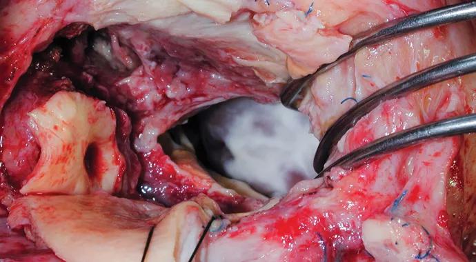
Image content: This image is available to view online.
View image online (https://assets.clevelandclinic.org/transform/fbcf8f3f-8a2f-4615-aae7-1b6930a64b52/Endocarditis-690x380_jpg)
Infective Endocarditis
A group of Cleveland Clinic specialists has created an atlas illustrating concepts essential to understanding infective endocarditis (IE), including its pathologic features, complications and surgical treatment.
Advertisement
Cleveland Clinic is a non-profit academic medical center. Advertising on our site helps support our mission. We do not endorse non-Cleveland Clinic products or services. Policy
Gosta B. Pettersson, MD, PhD, Vice Chairman of the Department of Thoracic and Cardiovascular Surgery in Cleveland Clinic’s Sydell and Arnold Miller Family Heart & Vascular Institute, led a group of cardiothoracic, infectious disease and quantitative health specialists in creating “Infective endocarditis: An atlas of disease progression for describing, staging, coding and understanding the pathology,” published in the April 2014 Journal of Thoracic and Cardiovascular Surgery (JTCS).
“There is so much misunderstanding of this disease, and only a limited number of surgeons have enough experience with endocarditis to really understand the pathology,” Dr. Pettersson says. “I have been in a position to, step-by-step, gain a deeper understanding of what I’m dealing with, witness the anatomy and interpret what is actually destroyed and not destroyed, and then determine what needs to be done, and how aggressive you can be — and need to be — to remove infected and necrotic tissue.”
At its core, the atlas is collection of observations and photographs from the operating room that illustrate key concepts of IE that Dr. Pettersson and his colleagues have developed over the years; these photos are supplemented by a more comprehensive online collection of images and echocardiographic videos.
“We have described our understanding of the pathophysiology and pathologic features of IE, translated this into an instrument for recording and coding, and illustrated the main pathologic concepts in an atlas,” Dr. Petterson and his team write in their JTCS article.
Advertisement
The aim was to provide a starting point for developing a standardized system for reporting the extent and pathologic features of IE to better understand and compare results of surgeries to treat IE. The intent is for this observation and documentation from the operating room to be combined with all the other information coded and collected — including the Society of Thoracic Surgeons’ Adult Cardiac Surgery Database — to create uniformity of reporting. Another byproduct: better correlation of pathologic findings with clinical presentation and other patient data.
This should ultimately help improve treatment strategies and the use and timing of surgery for optimal patient outcomes, Dr. Pettersson notes. “We can all take advantage of this work,” he says. “In this database we have carefully reviewed and coded the pathology of all surgical IE cases. Anyone interested in endocarditis who has a good idea for a study can take advantage of it. We have invested a lot of effort in creating this database, and we have been very successful in improving outcomes for these most difficult patients.”
The group’s findings indicate that the key to understanding IE is appreciating its pathologic progression: Circulating organisms or bacteria adhere to damaged areas of the endocardium, endothelium or foreign material in the bloodstream (such as prosthetic heart valves or pacemaker leads).
These damaged areas often have deposits of platelets, fibrin or clots that facilitate organism or bacteria growth. The organisms grow and produce a protective slime that attracts fibrin and cell deposits to form vegetations, a source of embolization. The organisms also produce enzymes and toxins that kill and disintegrate underlying tissue, resulting in leaky valves and invasion outside the circulation.
Advertisement
By understanding the pathology of IE, the group was able to develop a form for coding pathologic features and proposed an extensive coding schema that helps explain why endocarditis behaves the way it does. The atlas’ major concepts are as follows:
“The atlas helps you understand the basics of this disease — what it does and why, and how the complications that we see relate to the nature of the disease,” Dr. Pettersson explains. “The simplest things are still not understood by everybody.”
Advertisement
He adds: “This atlas is my personal, most important contribution to research in its simplicity.”
The next step is to dig deeper into this relatively rare disease. Dr. Pettersson notes he is just beginning to see the numbers that allow him to distinguish differences in subcategories of patients.
“This is a disease that requires contributions from infectious disease, cardiology, cardiac surgery and sometimes other specialties,” he says. “It is not only a locally destructive disease in the heart, but a serious disease with very serious complications. Overall, endocarditis is the most serious of all valve diseases.”
He and his colleagues are also moving forward on establishing a formal Endocarditis Center with a devoted group of physician specialists from infectious disease, cardiology and cardiac surgery that will focus on management plans and algorithms for patients from around the world with the most advanced cases of endocarditis.
Advertisement
Advertisement

A sampling of outcome and volume data from our Heart & Vascular Institute

Concomitant AF ablation and LAA occlusion strongly endorsed during elective heart surgery

Large retrospective study supports its addition to BAV repair toolbox at expert centers

Young age, solid tumor, high uptake on PET and KRAS mutation signal risk, suggest need for lobectomy

Surprise findings argue for caution about testosterone use in men at risk for fracture

Residual AR related to severe preoperative AR increases risk of progression, need for reoperation

Findings support emphasis on markers of frailty related to, but not dependent on, age

Provides option for patients previously deemed anatomically unsuitable