Locations:

With 7T MRI, an image is worth more than a thousand words
System-level brain study at a nearly microscopic scale. That’s what neuroscience researchers have been relishing since the installation of Cleveland Clinic’s 7-tesla (7T) MRI scanner in 2013. Here are a few glimpses of what it makes possible.
Advertisement
Cleveland Clinic is a non-profit academic medical center. Advertising on our site helps support our mission. We do not endorse non-Cleveland Clinic products or services. Policy
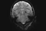
Image content: This image is available to view online.
View image online (https://assets.clevelandclinic.org/transform/74138035-3a3f-4a4b-81fc-8f9f97c42171/16-NEU-1680-Slide-1-150x103_jpg)
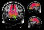
Image content: This image is available to view online.
View image online (https://assets.clevelandclinic.org/transform/117456e5-a232-4c76-9763-2f380b532cb8/16-NEU-1680-Slide-2-150x103_jpg)

Image content: This image is available to view online.
View image online (https://assets.clevelandclinic.org/transform/c3e83059-4f29-45ff-a6d5-9e4c34c2bac9/16-NEU-1680-Slide-3-150x103_jpg)
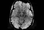
Image content: This image is available to view online.
View image online (https://assets.clevelandclinic.org/transform/b642b805-e874-4261-a36f-823047c47450/16-NEU-1680-Slide-4-150x103_jpg)
Advertisement
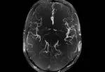
Image content: This image is available to view online.
View image online (https://assets.clevelandclinic.org/transform/2f252d85-5844-4539-a93f-91be68df3aba/16-NEU-1680-Slide-5-150x103_jpg)
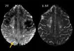
Image content: This image is available to view online.
View image online (https://assets.clevelandclinic.org/transform/3f13424e-9040-40a8-9d61-07196b1c7a14/16-NEU-1680-Slide-6-150x103_jpg)
Slide 1/6
Images were provided by Mark Lowe, PhD, Cleveland Clinic’s Director of High-Field MRI; Sehong Oh, PhD, project staff scientist in Cleveland Clinic’s Imaging Institute; and Stephen E. Jones, MD, PhD, Vice Chair for Research and Academic Affairs in the Imaging Institute.
RELATED: Ultra-High-Field MRI: A Snapshot of Its Growing Utility in Neuro Care
Advertisement
Advertisement

New study advances understanding of patient-defined goals

Testing options and therapies are expanding for this poorly understood sleep disorder

Real-world claims data and tissue culture studies set the stage for randomized clinical testing

Digital subtraction angiography remains central to assessment of ‘benign’ PMSAH

Cleveland Clinic neuromuscular specialist shares insights on AI in his field and beyond

Findings challenge dogma that microglia are exclusively destructive regardless of location in brain

Neurology is especially well positioned for opportunities to enhance clinical care and medical training

New review distills insights from studies over the past decade