Locations:

Researchers aim to stop visual scarring in the human eye
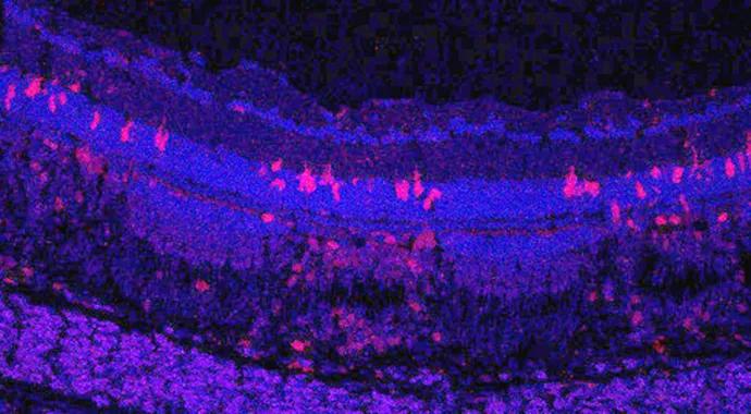
Image content: This image is available to view online.
View image online (https://assets.clevelandclinic.org/transform/bdc75ff3-6a27-4e3f-b5c3-3ceb31cf76b8/690x380-Yuan_jpg)
zebrafish
Zebrafish eyes are much like their counterparts in humans. But unlike humans, zebrafish can prevent scarring and regenerate damaged retinas.
Advertisement
Cleveland Clinic is a non-profit academic medical center. Advertising on our site helps support our mission. We do not endorse non-Cleveland Clinic products or services. Policy

Image content: This image is available to view online.
View image online (https://assets.clevelandclinic.org/transform/bd94bc82-4ab2-46bd-845a-c6302e0b49d8/jan2015-subscribe-cqdpulse3_jpg)
In the laboratory of ophthalmologist Alex Yuan, MD, PhD, at Cleveland Clinic’s Cole Eye Institute, scientists are using a zebrafish model to study how and why these processes take place, and how they might be used to prevent scar formation and stimulate retinal regeneration in the human eye.
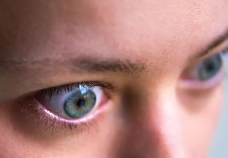
The human retina is subject to harm from many sources, including trauma, disease (i.e., macular degeneration and diabetic retinopathy), retinal tears or detachment, inherited disorders and other causes. It responds to different types of injury in a very similar way: when damaged, scars form.
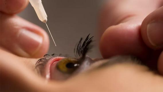
The ophthalmic laser is applied to the retina to treat existing retinal disease, such as macular edema, retinal tears, proliferative diabetic retinopathy, retinal vein occlusion, choroidal neovascularization and retinal tumors. The goal of most treatments is to stop disease progression and preserve or improve vision.
However, the application of lasers to the retina often causes loss of vision through an indirect adverse effect on neighboring healthy tissues. “Currently, laser treatment causes scotomas from areas where laser scars form. Over time, these scars can even enlarge, further jeopardizing sight,” says Dr. Yuan.
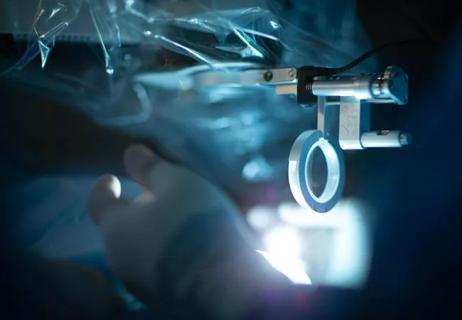
Traditionally, retinal injury in fish can be caused by intense light or chemicals, or through mechanical means. These methods, however, cause too much collateral damage. To avoid this drawback, Dr. Yuan’s laboratory is using a new laser injury model to uncover the cellular and molecular differences between the zebrafish and human injury response.
Advertisement
Zebrafish can be genetically manipulated to enable study of the effect of modifying different genes on the regeneration response. The fish are also a cone-dominated species, as are humans. Mice, however, are rod-dominant.
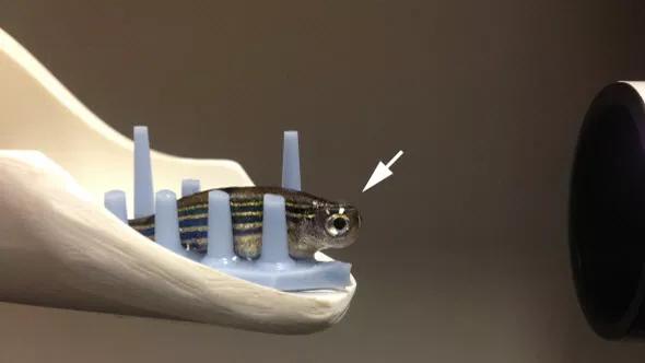
Image content: This image is available to view online.
View image online (https://assets.clevelandclinic.org/transform/73ca5b76-29d2-4b78-8dea-3459b7eb10ef/Fish_jpg)
Image of an adult zebrafish with a contact lens placed over the eye (arrow). Laser treatment is applied through this lens.
The zebrafish injury response is similar to that in humans. The same cell types are involved, but one of them (the Müller cell in zebrafish) turns into a progenitor or stem cell that stops scarring and promotes regeneration after retinal damage. In humans, this same cell forms a scar. “My lab is figuring out why this difference exists, and whether we can coax human Müller cells to respond differently and become progenitor cells,” says Dr. Yuan.
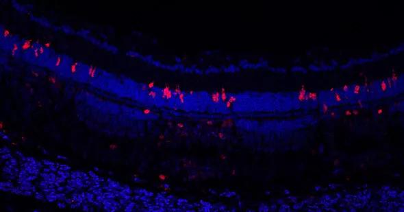
Image content: This image is available to view online.
View image online (https://assets.clevelandclinic.org/transform/b6d94247-c84b-45b6-8248-cb40caec3f07/Proliferating-cells_jpg)
Cryosection of a zebrafish retina, two days after laser lesions were placed. The blue labels individual nuclei and the red labels proliferating cells in the retina. The red cells are clustered over the three laser lesions seen in this image.
The main goals of Dr. Yuan’s laboratory are not just to study the response to laser injury, but to also learn how to prevent scar formation, and to stimulate retinal regeneration. “Learning how to limit scar formation or reverse it would be beneficial,” he says, “but being able to stop scar formation would have a tremendous impact.”
According to Dr. Yuan, scar formation is the leading reason for failure after retinal detachment repair. It’s also why patients with macular degeneration end up with poor vision. “If we can learn how to stop scar formation it would be a huge advance,” he says, “but if we can take the next step — to stop scar formation and regenerate the retina — that would be our ultimate dream.”
Advertisement
Advertisement
Early data shows risk is 73% higher in patients with lupus, 40% higher in patients with rheumatoid arthritis

Identifies weak spots in the cornea before shape change occurs
Study highlights the value of quantitative ultra-widefield angiography
Switching medications may decrease treatment burden and macular fluid
Interventions abound for active and stable phases of TED
Corneal imaging and interpretation play a major role
Cole Eye Institute imaging specialists are equal parts technician, artist and diagnostician

Effect of low-dose atropine and dual-focus contact lenses is unknown in patients with comorbid eye conditions