Locations:

Promises a low-cost way to visualize areas of stenosis
A 3D-printed model of an atherosclerotic superficial femoral artery (SFA) can be used to provide realistic-appearing ultrasound characteristics at very low cost. So concludes a recent study by Paul Bishop, MSEE, RVT, and his colleagues in Cleveland Clinic’s Department of Vascular Surgery and Department of Biomedical Engineering.
Advertisement
Cleveland Clinic is a non-profit academic medical center. Advertising on our site helps support our mission. We do not endorse non-Cleveland Clinic products or services. Policy
Using commercially available 3D printing materials and equipment, the researchers created a 3D model of an atherosclerotic SFA based on actual geometry derived from a CT scan reconstructed and segmented using semi-automated methods and commercial software. Multiple 3D print materials were selected to simulate normal artery wall tissue and atherosclerotic plaque. When the researchers evaluated the 3D-printed model with ultrasound, they demonstrated that lumen geometry of the SFA model was similar to the geometry of the actual artery. Ultrasound was able to discern between the 3D-printed materials and to visualize regions with stenosis, as shown in the sample images below.
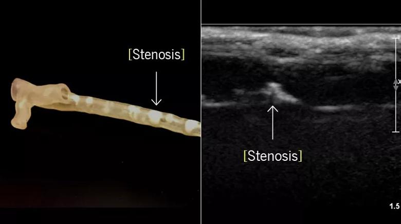
Image content: This image is available to view online.
View image online (https://assets.clevelandclinic.org/transform/683b80c2-f2fc-4464-95f4-bba457b37a18/18-HRT-4695-3D-printed-SFA-CQD-Inset_jpg)
Stenosis in a 3D-printed superficial femoral artery model (left) is matched with its corresponding appearance on ultrasound (right).
Imaging replication was not perfect, however: Ultrasound measures of echogenicity and wave velocity were noted to differ between the model and biological tissue.
“Although the 3D-printed model didn’t demonstrate fully accurate ultrasound characteristics, it provided realistic imaging on our first attempt to create an ultrasound phantom using only commercially available equipment and materials,” says Bishop, Director of Cleveland Clinic’s Vascular Core Laboratory. “Visualization of the SFA model wall was enabled much as would be the case with an in vivo SFA despite differences in ultrasound properties from actual tissue.”
While noting that further research is needed to refine 3D printing materials to better replicate biological tissue, Bishop and colleagues say their model may be useful in cost-sensitive applications in which exact ultrasound accuracy is not necessary. Indeed, they estimate their total 3D printing material cost for the model to be under $20.
Advertisement
Their study was awarded the D.E. Strandness, MD, Scientific Award for Excellence in Scientific Research from the Society for Vascular Ultrasound in 2017 and has been submitted for publication.
Contact Bishop at bishopp@ccf.org.
Advertisement
Advertisement

A sampling of outcome and volume data from our Heart & Vascular Institute

Concomitant AF ablation and LAA occlusion strongly endorsed during elective heart surgery

Large retrospective study supports its addition to BAV repair toolbox at expert centers

Young age, solid tumor, high uptake on PET and KRAS mutation signal risk, suggest need for lobectomy

Surprise findings argue for caution about testosterone use in men at risk for fracture

Residual AR related to severe preoperative AR increases risk of progression, need for reoperation

Findings support emphasis on markers of frailty related to, but not dependent on, age

Provides option for patients previously deemed anatomically unsuitable